Crohn disease


Crohn disease, also known as regional enteritis, is an idiopathic inflammatory bowel disease characterized by widespread discontinuous gastrointestinal tract inflammation. The terminal ileum and proximal colon are most often affected. Extraintestinal disease is common.
Epidemiology
The diagnosis is typically made between the ages of 15 and 25 years with no gender predilection . There is a familial component and incidence varies geographically.
Clinical presentation
Patients typically present with chronic diarrhea and recurrent abdominal pain, although occasionally the presentation is with a complication or an extraintestinal manifestation. Anemia may be present and C-reactive protein may be elevated .
Fecal calprotectin has been increasingly used in recent years to:
- distinguish inflammatory bowel disease from irritable bowel syndrome
- assess disease activity in inflammatory bowel disease, including acute exacerbations and response to treatment
Pathology
Crohn disease remains idiopathic, although infective agents have been gaining in popularity as a possible cause, including the measles virus and atypical mycobacterium. As there are definite genetic factors at play, multiple factors are likely to contribute . Incidence is higher in people with first degree relatives having IBD, reaching up to 10%. Also, there has been shown 30-50% chance of developing the disease in mono- or heterozygous twins.
Initially, the disease is limited to the mucosa with neutrophilic cryptitis and lymphoid hyperplasia, lymphedema and shallow aphthoid ulceration. As the disease progresses, the entire bowel wall becomes involved, with linear longitudinal and circumferential ulcers extending deep into the bowel wall, predisposing to fistulae. Inflammation also extends into the mesentery and over time leads to chronic fibrotic change, and stricture formation .
Inflammation can occur anywhere along the digestive tract, including the mouth and esophagus. Crohn disease of the esophagus should only be considered if Crohn disease of the bowel is already evident . The following findings are evidences of Crohn disease involving the mouth and esophagus :
- mucogingivitis mucosal tags, deep ulcerations, cobblestoning, lip swelling and pyostomatitis vegetans, esophageal ulcers and strictures
Extraintestinal manifestations include :
- skin
- erythema nodosum
- pyoderma gangrenosum
- aphthous stomatitis
- enterocutaneous fistulas
- joints
- arthritis
- seronegative spondyloarthritis
- sacroiliitis (one of the most frequent extraintestinal manifestations)
- eyes
- episcleritis
- iritis
- uveitis (acute anterior uveitis)
- liver and biliary system
- pericholangitis
- primary sclerosing cholangitis (PSC) (more common in ulcerative colitis)
- autoimmune hepatitis
- cirrhosis
- gallstones: seen in 30-50%
- hepatic abscess
- pancreatitis
- acute cholecystitis
- portal vein thrombosis (rare, yet higher than the general population)
- fatty infiltration of the liver (steroid therapy, hyperalimentation)
- renal tract
- renal calculi containing oxalate
- the poor fat absorption results in binding of calcium by fats, which in turn reduces the amount of calcium that can bind to oxalate, therefore increasing the amount of unbound oxalate available for resorption; this resorption occurs in the colon, and therefore patients with an ileostomy do not have the same increased risk
- ileoureteral / ileovesical fistula
- renal calculi containing oxalate
- pulmonary and thoracic associations
Classification
Recognized classifications include:
- Montreal classification of inflammatory bowel disease
- Vienna classification of inflammatory bowel disease
Radiographic features
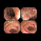
The characteristic of Crohn disease is the presence of skip lesions and discrete ulcers. The frequency with which various parts of the gastrointestinal tract are affected varies widely :
- small bowel: 70-80% ; the terminal ileum is usually affected first
- small and large bowel: 50%
- large bowel only: 15-20%
The choice of investigation modality depends on local expertise and availability. CT and MR enterography are similar in sensitivity for active inflammation (89% vs 83% respectively) and both are better than small bowel follow-through (67-72%) . The lack of ionizing radiation from MRI would make it a better option, however, the availability of MRI is limited in many countries.
Ultrasound, which has been largely applied by gastroenterologists, is also an option for diagnosing active disease, following-up treatment response, and assessing complications . Reported sensitivity of 75-94% and specificity 67-100% .
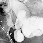
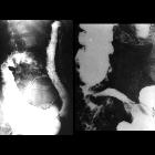
Fluoroscopy
Features on barium small bowel follow-through include:
- mucosal ulcers
- aphthous ulcers initially
- deep ulcers (>3 mm depth)
- longitudinal fissures
- transverse stripes
- when severe leads to cobblestone appearance
- may lead to sinus tracts and fistulae
- widely separated loops of bowel due to fibrofatty proliferation (creeping fat)
- thickened folds due to edema
- pseudodiverticula/pseudosacculation formation: due to contraction at the site of ulcer with ballooning of the opposite (usually antimesenteric) site
- string sign: tubular narrowing due to spasm or stricture depending on the chronicity
- partial obstruction
- on control films presence of gallstones, renal oxalate stones, and sacroiliac joint or lumbosacral spine changes should be sought
Ultrasound
Ultrasound has a limited role, but due to it being cheap, available, and not involving ionizing radiation, it has been evaluated as an initial screening tool for active disease and also for follow-up and to assess complications . Features which may be discovered during transabdominal sonography include :
- small bowel wall thickening (>3-4 mm)
- needs to be interpreted in the context of pretest probability
- affected loops often non-compressible and are difficult to displace with transducer pressure
- affected segments lose peristaltic activity
- loss of mural stratification
- the gut signature is characteristic of small bowel
- chronic disease activity may result in collagen deposition within the wall, which imitates the appearance of the normal submucosa
- bowel wall hyperemia
- hyperechoic, circumferential layer external to the bowel wall
- represents fibrofatty proliferation, thought to represent active inflammation
- interruption by hypoechoic mesenteric streaks imply a greater degree of inflammation
- mesenteric lymphadenopathy
- three or more mesenteric lymph nodes enlarged, measured over 4 mm (width) or 8 mm (length)
- free intraperitoneal fluid
- thought to be secondary to transmural inflammation
On pulsed-wave Doppler evaluation, increased superior mesenteric artery (SMA) flow volume and decreased SMA resistive index (SMA RI) also correlate with disease activity. Successful treatment may result in the normalization of these imaging parameters .
Complications may also be assessed with sonography, with the following features commonly detected in the following :
- abscess formation
- irregular, echo-poor collection with a variable amount of internal echogenic collections and posterior acoustic enhancement
- absence of internal flow (by color flow Doppler)
- may demonstrate peripheral vascularity with power Doppler
- fistula creation
- linear, anechoic tract extending from an affected bowel loop to another structure, often with scattered hyperechoic puncta with inhomogeneous posterior acoustic shadowing (air)
- diameter of the fistulous tract should be <2 cm
- luminal stenosis
- segmental wall thickening, loss of peristalsis, luminal narrowing or obliteration, preceding segment dilated (>2.5 cm)
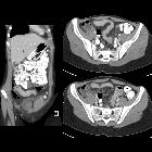
CT
CT is commonly the first imaging assessment of those patients in the setting of acute abdomen, or it can be also applied to the reassessment of complications in patients with known Crohn disease. Common features include:
- mural hyperenhancement
- fat halo sign: submucosal fat deposition
- bowel wall thickening (1-2 cm), which is most frequently seen in the terminal ileum (present in up to 83% of patients)
- comb sign: engorgement of the vasa recta
- perienteric fat stranding
- affected bowel loops separated by focal/regionally increased fat (fibrofatty proliferation; creeping fat)
- strictures and fistulae, with upstream dilatation
- mesenteric/intra-abdominal abscess or phlegmon formation
- abscesses are eventually seen in 15-20% of patients
CT is also able to give valuable information on:
- perianal disease
- hepatobiliary disease
CT enterography
CT enterography is superior to standard CT studies when assessing for small bowel Crohn disease and is equivalent to MR enterography, the downside compared to the latter being ionizing radiation. CT enteroclysis may be attempted in select patients. For the imaging findings and descriptors in CT enterography, see the discussion below under MR enterography.
MRI
MR enterography has become an increasingly important part of the management of patients with Crohn disease. MR enteroclysis may be attempted in select patients.
MR enterography (MRE)
Features of small bowel Crohn disease to be assessed on dedicated MRI study include:
- segmental mural hyperenhancement
- asymmetric
- specific sign for Crohn disease
- tends to more frequently involve the mesenteric border of a small bowel loop
- stratified: layers of enhancement within the bowel wall
- bilaminar: hyperenhancement of the inner layer
- please note that the term “mucosal hyperenhancement” is discouraged given the mucosa itself is absent in the Crohn inflamed bowel segments
- trilaminar: hyperenhancement of both the inner and outer layers of the small bowel wall
- more frequently seen on MR enterography (cf. CT enterography)
- it can be explained by a combination of intramural submucosal edema, granulation tissue, intramural fat deposition (fat halo sign on CT), or fibrosis
- bilaminar: hyperenhancement of the inner layer
- homogeneous or transmural hyperenhancement: involving the entire small bowel wall
- less specific and, depending on distribution, other differentials should apply
- asymmetric
- DWI: restrict diffusion is seen within the small bowel wall segments of active inflammation
- wall thickening : must be assessed in a loop adequately dissented by fluid
- mild 3-5 mm
- moderate >5-9 mm
- severe > 9mm
- please note that focal thickening over 15 mm is atypical for Crohn, and consideration for malignancy should be sought
- intramural edema
- better assessed on MRI T2 fat-saturated or low b-value DWI sequences
- increased T2 signal in the thickened bowel wall is particularly helpful in evaluating for acute inflammation
- stricture: focal narrow of the lumen with immediate upstream bowel dilatation >=3 cm. Bear in mind that sometimes the upstream loop is not dilated to size criterion due to decompression by a fistula/penetrating disease or limited bowel content inflow in the setting of two or more consecutive stricture segments
- highly suspected stricture should be reported in persistent focal luminal narrowing across multiple MRI sequences
- although they can be divided as fibrotic or inflamed strictures, the majority will have a component of active inflammation
- location and length of the stricture should be reported
- strong association of strictures and penetrating disease, so one should prompt the evaluation for the other
- stricture vs obstruction: upstream dilatation over 4 cm is advised to be reported as small bowel obstruction
- ulcerations: focal defect at the intraluminal aspect of the small bowel wall, confined within the serosa (cf. sinus tract)
- decreased peristalsis: coronal cine MRI sequences (bSSFP) can also be useful in diagnosis as inflamed loops of bowel frequently demonstrate decreased peristalsis
- pseudodiverticula/pseudosacculation formation: broadband outpouching along the antimesenteric border
Mesenteric signs of inflammation include:
- engorged vasa recta: enlarged mesenteric blood vessels related to an inflamed bowel loop (comb sign)
- fibrofatty proliferation (creeping fat):
- hypertrophy of the mesenteric fat, which separates bowel loops
- mesenteric venous thrombosis
- usually adjacent to an inflamed loop
- acute: distended vein
- chronic: narrowed or interrupted vein
- lymphadenopathy
Features of penetrating disease include:
- sinus tract: blind-ended tract extending beyond the bowel serosa
- fistula tract: tract that communicates the bowel lumen to another epithelialize surface
- usually seen proximal to a small bowel stricture
- active inflammation is almost always present
- complex fistula: more than one tract associated with angulation or tethering of affected loops
- inflammatory mass: mesenteric stranding (on CT) or increased T2 signal denoting inflammation but without fluid collections
- abscess: a fluid collection with rim-enhancement
Extraintestinal involvement can be at least partially assessed on MR enterography, particularly, hepatobiliary disease (eg. gallstones) and sacroiliitis.
MRI pelvis
Crohn perianal disease is mostly characterized on imaging by perianal fistulas and is better evaluated using a dedicated MRI protocol for the anal canal.
Treatment and prognosis
Management is complex as the condition is chronic with a relapsing-remitting course. Medical management includes corticosteroids, 5-ASA preparations, immunomodulation (e.g. azathioprine, cyclosporine, methotrexate) . Surgical management is reserved for complications including:
- strictures
- adhesions and bowel obstructions
- fistulae
- perianal disease
History and etymology
It is named after Burrill Bernard Crohn (1884-1983), an American gastroenterologist, who described the condition as 'regional ileitis' in his seminal 1932 paper . However, the first definite description (but see below) was nearly twenty years prior, by Sir T (Thomas) Kennedy Dalziel (1861-1924), a Scottish surgeon, in 1913 .
Antoni Leśniowski (1867-1940), a Polish surgeon, described a small bowel condition in 1904 in a small series of four patients, with similarities to Crohn disease, although it remains controversial if it was actually Crohn's . At least one of the patients probably actually had ileal tuberculosis. Nevertheless, Polish physicians and journals usually call the condition Lesniowski-Crohn disease.
Differential diagnosis
The differential diagnosis depends on the presenting symptom. When terminal ileitis is the main presentation, then differentials (adjusted for patient's age) include :
- acute appendicitis
- Yersinia ileitis
- mesenteric adenitis
- ileocecal tuberculosis
- malignancy
When colonic involvement is the predominant feature then other considerations include:


 Assoziationen und Differentialdiagnosen zu Morbus Crohn: