masticator space
The masticator space is one of the deep compartments of the head and neck.
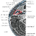
Gross anatomy
The masticator space are paired suprahyoid cervical spaces on each side of the face. Each space is enveloped by the superficial (investing) layer of the deep cervical fascia. The superficial layer of deep cervical fascia splits into two at the lower border of the mandible. The inner layer runs deep to the medial pterygoid muscle and attaches to the skull base medial to foramen ovale. The outer layer covers the masseter and temporalis muscles and attaches to the parietal calvaria superiorly.
Contents
- muscles of mastication
- ramus and body of mandible
- mandibular division of the trigeminal nerve (Vc)
- enters the masticator space via the foramen ovale
- inferior alveolar nerve
- inferior alveolar artery and vein
- pterygoid venous plexus
Boundaries and relations
- anteriorly: buccal space
- posterolaterally: parotid space
- medially: parapharyngeal space
Communications
Masticator space malignancy or infection can spread perineurally via the foramen ovale and along the course of the mandibular division of the trigeminal nerve into the middle cranial fossa.
Radiographic appearance
Ultrasound
- limited use when imaging the masticator space
- masseter muscles, zygomatic arch, outer cortex of the ramus of mandible and suprazygomatic segment of temporal muscle can all be visualized
- limited visualization of a number of important structures :
- pterygoid muscles
- pterygoid venous plexus
- mandibular branch of the trigeminal nerve
CT
- best modality for detecting bony erosion in the cortex of the mandible and is excellent for characterizing tumor matrix mineralization
- abscess in the masticator space shows up as a fluid collection with peripheral rim enhancement whereas a phlegmon shows low density edematous tissue without peripheral enhancement
- schwannoma appears as a well-circumscribed fusiform mass with extension through the foramen ovale and is higher in attenuation that adjacent muscle and shows contrast enhancement
MRI
MRI better characterizes soft tissue invasion by tumors and perineural tumor spread :
- schwannoma demonstrates intermediate signal on T1-WI and hyperintensity on T2-WI with contrast enhancement
- neurofibroma generally show heterogeneity on T2-WI and heterogeneous contrast enhancement
- locally invasive carcinoma from the nasopharynx or oral cavity demonstrate intermediate-to-high signal on T2-WI and low signal on T1-WI, with or without bone destruction and perineural spread; lymphadenopathy is common
Nuclear medicine
- PET-CT with fluorine-18 FDG can be used to detect metastatic disease and to differentiate recurrent tumors from post-radiation change
Related pathology
- odontogenic abscess
- osteomyelitis
- direct spread of squamous cell carcinoma
- lymphoma
- minor salivary gland tumors
- muscle sarcoma
- bone sarcoma
- osteoradionecrosis
- schwannoma
- neurofibroma
- benign masseteric hypertrophy
- accessory parotid tissue
Siehe auch:
- Osteomyelitis
- carotid space
- parotid space
- deep compartments of the head and neck
- muscles of mastication
- Plattenepithelkarzinom
- Retropharyngealraum
- superficial mucosal space
- prevertebral space
- Mandibula
- Parapharyngealraum
- fascial spaces of the head and neck
und weiter:


 Assoziationen und Differentialdiagnosen zu masticator space: