trigeminal nerve


The trigeminal nerve is the fifth cranial nerve and its primary role is relaying sensory information from the face and head, although it does provide motor control to the muscles of mastication. It is both large and complicated and has multiple brainstem nuclei (sensory and motor) as well as many interconnections with other cranial nerves. It swaps parasympathetic fibers and taste fibers somewhat haphazardly and divides into numerous terminal branches.
Gross anatomy
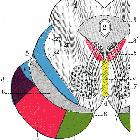
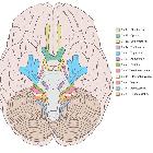
Nuclei
There are four cranial nerve nuclei: three sensory and one motor.
The sensory nuclei are arranged in a column which spans from the midbrain through the pons and medulla and into the upper cervical cord.
The motor nucleus is located in the upper pons and gives off the smaller motor root which bypasses the trigeminal ganglion and innervates the muscles of mastication as well as mylohyoid, the anterior belly of digastric, tensor tympani and tensor palatini.
Intracranial component
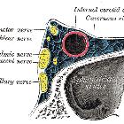
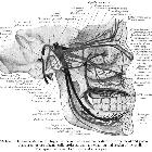
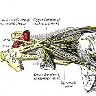
The trigeminal nerve exits at the mid pons anteriorly, courses through the prepontine cistern (cisternal portion), and crosses the porus trigeminus to enter a prolongation of dura at the apex of the petrous temporal bone known as the Meckel cave (cavernous portion) where its fibers form the trigeminal ganglion, which is also known as the Gasserian or semilunar ganglion. It then divides into three main branches known as divisions:
Ophthalmic division / nerve (V1 or Va)
Courses anteriorly in the lateral wall of the cavernous sinus inferior to the trochlear nerve and is crossed medially by the oculomotor nerve. Just before entering the orbit, the tentorial nerve arises and ascends to supply a large portion of the falx and supratentorial dura. The ophthalmic division then divides into 3 terminal branches before each passes through the superior orbital fissure separately:
- frontal nerve (superior orbital fissure outside the tendinous ring)
- lacrimal nerve (superior orbital fissure outside the tendinous ring)
- nasociliary nerve (superior orbital fissure through the tendinous ring)
Maxillary division / nerve (V2 or Vb)
Courses anteriorly low in the lateral wall of the cavernous sinus inferior to the ophthalmic division. Just before exiting the skull it runs along the floor of the middle cranial fossa and gives off the middle meningeal nerve which ascends to supply the anterior dura of the middle cranial fossa. It then passes through the foramen rotundum in the greater wing of the sphenoid bone to exit the skull and enter the superior aspect of the pterygopalatine fossa. It gives branches to the pterygopalatine ganglion but also receives parasympathetic nerves from the ganglion via the greater petrosal nerve. It then divides into the:
- zygomatic nerve (inferior orbital fissure)
- infraorbital nerve (inferior orbital fissure then infraorbital foramen)
- nasopalatine nerve (sphenopalatine foramen)
- posterior superior nasal nerves (sphenopalatine foramen)
- greater palatine nerve (greater palatine foramen)
- lateral posterior inferior nasal nerve (un-named foramina)
- lesser palatine nerve (lesser palatine foramen)
- pharyngeal nerve (palatovaginal canal)
Mandibular division / nerve (V3 or Vc)
Courses inferiorly through the foramen ovale to enter the infratemporal fossa, hence it does not pass through the cavernous sinus. It consists of a sensory root and a smaller motor root, the latter which bypasses the trigeminal ganglion inferiorly. These roots pass through the foramen ovale separately and then unite just below the foramen. It immediately gives off nervus spinosus and nerve to medial pterygoid from the main trunk. It then descends in the infratemporal fossa passing between the tensor veli palatini and lateral pterygoid muscles before dividing into anterior and posterior divisions:
- anterior division (4 branches, all motor except one)
- posterior division (3 branches, all sensory except one)
Related pathology
Siehe auch:
- Pons
- Fissura orbitalis superior
- Mesencephalon
- Hirnnerven
- trigeminal schwannoma
- Anatomie Hirnstamm
- muscles of mastication
- Ganglion Gasseri
- Orbita
- Nervus ophthalmicus
- Nervus maxillaris
- Nervus mandibularis
und weiter:
- Nervus facialis
- Sinus cavernosus
- Arteria trigemina
- trigeminal nerve branches (mnemonic)
- Gradenigo-Syndrom
- inferolateral trunk
- Cavum trigeminale
- Musculus digastricus
- mandibular foramen
- vertebrobasiläre Dolichoektasie
- Nervus nasopalatinus
- Plexus cervicalis
- cavernous sinus (mnemonic)
- mandibular nerve
- Pyramidenspitzen-Osteomyelitis
- Hirnnerven Eselsbrücke
- Nucleus mesencephalicus nervi trigemini
- Parapharyngealraum


 Assoziationen und Differentialdiagnosen zu Nervus trigeminus: