parotid gland


The parotid gland is the largest of the salivary glands and secretes saliva via the parotid duct into the oral cavity to facilitate mastication and swallowing. It is located in the parotid space.
Gross anatomy
The parotid gland is wrapped around the mandibular ramus and extends to a position anterior and inferior to the ear. It has superficial and deep lobes, separated by the facial nerve.
The facial nerve and its branches pass through the parotid gland, as does the external carotid artery and retromandibular vein. The external carotid artery forms its two terminal branches within the parotid gland: maxillary and superficial temporal artery.
The gland usually contains several intraparotid lymph nodes. These lymph nodes are the first station of lymphatic drainage of the skin of the pinna and peri-auricular skin.
A fibrous capsule surrounds the gland, formed by the superficial (investing) layer of the deep cervical fascia, creating the parotid space. Posteriorly, this fascia condenses to form the stylomandibular ligament.
The inferior projection of the parotid is often referred to as the "tail", which overlies the angle of the mandible. The tail is not distinct from the rest of the gland, but it has been defined as the inferior 2 cm of the gland .
Anteriorly, there is often an accessory parotid gland, which may be separate from the main gland.
There is fatty infiltration or fatty replacement of the parotid glands with age .
Relations
- superior pole: external acoustic meatus, temporomandibular joint
- lower pole: behind the angle of the mandible, anterior to the sternocleidomastoid and posterior belly of the digastric
- lateral surface: subcutaneous tissue
- anterior surface: clasps the ramus of the mandible with the masseter on its outer surface and medial pterygoid on its inner surface inferiorly (separated by the stylomandibular ligament)
- anterior border: formed by the lateral edge of the anterior surface where it meets the masseter
- the parotid duct and five facial nerve branches emerge from this border
- from the deeper part, the superficial temporal and maxillary arteries leave the gland
- deep surface: indented by the mastoid process and its attached muscles (sternocleidomastoid and posterior belly of the digastric), styloid process and its attached muscles (stylohyoid, styloglossus, stylopharyngeus) and two ligaments (stylomandibular, stylohyoid)
- the external carotid artery enters the gland through this surface
- the styloid process separates the gland from the internal jugular vein and internal carotid artery
- the temporozygomatic and cervicofacial branches of the facial nerve enter the gland between the mastoid and styloid processes
Blood supply
- arterial: external carotid artery and a specific branch of the artery, the transverse facial artery
- venous drainage: plexus of veins into the internal jugular vein
Lymphatic drainage
Intraparotid nodes drain into the deep cervical chain.
Innervation
- sensory: auriculotemporal nerve, greater auricular nerve
- parasympathetic: via auriculotemporal nerve
- sympathetic: via plexus surrounding external carotid artery from superior cervical ganglion.
Variant anatomy
- accessory parotid gland
- facial process: anterior extension of glandular tissue along the parotid duct continuous with the main gland
- ectopic parotid tissue
- parotid duct duplication
- congenital agenesis: either unilateral and bilateral
Radiographic appearance
Ultrasoundis often the first diagnostic procedure to evaluate morphological and structural changes of the parotid gland; for small (<3 cm) and superficial lesions, ultrasound and cytology are often sufficient for a definitive diagnosis .
Ultrasound
- appears homogeneous with increased echogenicity compared to nearby muscle
- intraparotid lymph nodes are normally seen (unlike the submandibular gland)
- retromandibular vein and external carotid artery are also easily seen and by inference the facial nerve, which lies lateral to these vessels
- limitations of ultrasound are:
- difficulty visualizing deep lesions: the deep lobe is not able to be assessed as it is protected by the mandibular ramus
- difficulty visualizing deep extension
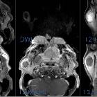
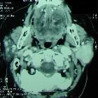
CT/MRI
- CT and MRI provide useful additional diagnostic imaging if malignancy is suspected , with the sensitivity approaching 100% for detecting parotid neoplasms
- the parotid duct and retromandibular vein are usually seen and approximate the plane separating the superficial and deep lobes
Related pathology
Siehe auch:
- Arteria carotis externa
- Nervus facialis
- pleomorphes Adenom der Glandula parotis
- Warthin-Tumor
- Tumoren der Speicheldrüsen
- sublingual gland
- Glandula submandibularis
- zystische Läsionen der Glandula parotis
- salivary glands
- major salivary glands
- minor salivary glands
- Vergrößerung der Glandula parotis
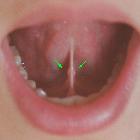
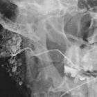
- Ductus parotideus
- Papilla parotidea
- anatomy of the head and neck
- Ramus mandibulae
und weiter:
- facial nerve branches (mnemonic)
- Sialolithiasis
- parotid space
- Arteria maxillaris
- Franceschetti-Zwahlen-Syndrom
- Pes anserinus
- ducts of the salivary glands
- posterior auricular artery
- Nervus petrosus minor
- foramen tympanicum
- myoepithelioma
- akzessorische Glandula parotis
- balloon dilatation of parotid duct stricture
- Pfeffer und Salz Erscheinungsbild
- parotid duct strictures
- MRI of the parotid glands in a patient with Sjogren's syndrome
- Jacobson-Anastomose


 Assoziationen und Differentialdiagnosen zu Glandula parotidea: