Sialadenitis


Sialadenitis refers to inflammation of the salivary glands. It may be acute or chronic and has a broad range of causes.
Clinical presentation
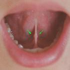
Patients may present with painful swelling of the concerned salivary gland, after eating (salivary colic). In bacterial sialadenitis, there may be a purulent discharge.
Pathology
Etiology
Sialadenitis can occur in various forms ranging from acute bacterial sialadenitis (acute suppurative sialadenitis) to acute viral sialadenitis to chronic sialadenitis.
Acute sialadenitis is most commonly caused by an ascending bacterial infection, with Staphylococcus aureus or Streptococcus viridans being the most common organisms . Sialolithiasis is often present (causing obstructive sialadenitis) and stones are found in ~85% of submandibular ducts and ~15% of parotid ducts . Other causes of acute sialadenitis include dehydration, immunosuppression, iatrogenic (drug-induced) and rarely haematogenous spread . Rarely sialadenitis may be secondary to an obstructive salivary duct carcinoma.
Epidemic parotitis is associated with the mumps virus, occurs mostly in children and is usually bilateral .
Associations
Other conditions related to sialadenitis include:
- Sjogren syndrome: chronic sialadenitis
- Mikulicz syndrome: chronic sialadenitis
- post-radiation: post-irradiative sialadenitis
- iodine-131 administration
- HIV-associated sialadenitis or immune reconstitution inflammatory syndrome (IRIS), often presenting with bilateral parotid swelling .
Distribution
Due to calculi being a dominant etiological factor, the distribution of sialadenitis follows that of sialolithiasis and therefore the submandibular glands are the most commonly affected (approximately 85%) as the submandibular ducts have more of an upward course and the secreted saliva has a higher viscosity .
Radiographic features
Fluoroscopy
Sialography is contraindicated in acute sialadenitis because it can worsen the infection .
Ultrasound
In acute sialadenitis, the affected gland appears enlarged, hypoechoic and hyperemic on ultrasound .
In chronic infective forms, the affected gland appears atrophic and diffusely hypoechoic with irregular margins - the ultrasound appearances have been likened to that of a “cirrhotic” liver .
There may be evidence of sialectasis if recurrent.
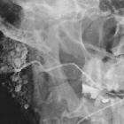
CT
Acute sialadenitis:
- enlarged salivary gland with abnormal attenuation, indistinct margin and vivid contrast enhancement with associated adjacent fat stranding and/or thickening of the deep cervical fasciathat is typically unilateral
- dilated duct from sialolithiasis or stenosis
- enlarged intra- or extra-glandular lymph nodes may also be seen but this is non-specific and can occur in other conditions such as malignancy
- abscesses are hypodense fluid collections, which may or may not be loculated
MRI
The salivary gland(s) is often enlarged. The affected gland can range from well-defined to poorly defined. Signal characteristics in the majority of cases tend to be heterogeneous .
Signal characteristics include:
- T1
- acute sialadenitis: low signal
- chronic sialadenitis: inhomogeneous low signal
- T2
- acute sialadenitis: overall signal tends to be high
- chronic sialadenitis: overall signal may be low-to-intermediate due to fibrosis
Treatment and prognosis
Treatment is usually endoscopic or surgical stone removal . If stone removal is unsuccessful the duct and gland may need to be removed if symptoms do not resolve.
Differential diagnosis
Differential conditions to keep in mind include :
See also
Siehe auch:
- Sialolithiasis
- Sjögren-Syndrom
- Glandula submandibularis
- sialosis
- chronic sclerosing sialadenitis
- küttner tumour
- benigne lymphoepitheliale Läsionen
und weiter:


 Assoziationen und Differentialdiagnosen zu Sialadenitis: