progressive massive fibrosis
Progressive massive fibrosis (PMF) refers to the formation of large mass-like conglomerates, predominantly in the upper pulmonary lobes, associated with radiating strands. These classically develop in the context of certain pneumoconioses (especially coal worker's pneumoconiosis and silicosis) although similar mass-like densities have occasionally been described with talcosis.
Radiographic features
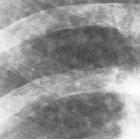
Plain radiograph
May be seen as large symmetric bilateral opacities with irregular margins in the upper lobes .
CT
Mass-like areas of lung opacification associated with radiating strands are seen; the "sausage-shaped" mass is characteristic. These regions commonly contain air bronchograms and calcifications . These areas can shrink over time and migrate towards the hilar regions .
MRI
Magnetic resonance imaging can be helpful for distinguishing between progressive massive fibrosis and lung cancer . The latter typically appears as T2-bright, whereas progressive massive fibrosis appears as T2-dark (compared to skeletal muscle) .
The most frequent MRI appearance are regions which have following signal characteristics :
- T1: iso- to hyperintense
- T2:
- hypointense (compared with skeletal muscle)
- areas of internal high T2 signal
- there may be rim enhancement
Nuclear medicine
On PET-CT, progressive massive fibrosis can be FDG-avid .
Differential diagnosis
Possible differential considerations include:
- pulmonary talc granulomatosis
- sarcoidosis
- lung cancer: has a higher SUVmax on PET-CT
In some situations consider pulmonary manifestations of sarcoidosis.
Siehe auch:
und weiter:


 Assoziationen und Differentialdiagnosen zu progressive massive Fibrose: