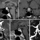
zystische Läsionen in der Hypophyse

MRI of a
benign pituitary cyst. Pituitary cysts are not rare and if large enough they can cause symptoms by compression of the pituitary gland or suprasellar structures.

Magnetic
resonance imaging of sellar and juxtasellar abnormalities in the paediatric population: an imaging review. Pars intermedia cyst. Sagittal T1WI (a) and axial T2WI (b) shows a small cyst between the anterior and posterior pituitary which lacks contrast enhancement on axial fat-suppressed T1WI (c)

Magnetic
resonance imaging of sellar and juxtasellar abnormalities in the paediatric population: an imaging review. Pituitary adenomas. Sagittal post-contrast fat-suppressed T1WI (a) shows an heterogenously enhancing pituitary macroadenoma which contains a non-enhancing portion with high signal on T2WI (b). secondary to cystic degeneration. Coronal post-contrast fat-suppressed T1WI (c) shows a pituitary microadenoma which shows less early contrast enhancement than the normal pituitary tissue
zystische Läsionen in der Hypophyse
Siehe auch:
- Rathke Zyste
- Tumoren der Hypophysenregion
- Empty-Sella-Syndrom
- verdickter Hypophysenstiel
- Kraniopharyngeom
- epidermale Inklusionszyste
- pituitary region mass with intrinsic high T1 signal
- Pars intermedia-Zyste der Hypophyse
- pituitary MRI - an approach
- mixed cystic and solid pituitary region mass
- purely intrasellar pituitary mass
- zystische Läsionen der Sellaregion
- supraselläre Arachnoidalzyste
- zystisches Hypophysenadenom
- SATCHMO
- solid and enhancing pituitary region masses
- cystic suprasellar mass

 Assoziationen und Differentialdiagnosen zu zystische Läsionen in der Hypophyse:


