zystische Läsionen der Sellaregion

MRI of a
benign pituitary cyst. Pituitary cysts are not rare and if large enough they can cause symptoms by compression of the pituitary gland or suprasellar structures.

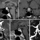
Magnetic
resonance imaging of sellar and juxtasellar abnormalities in the paediatric population: an imaging review. Pars intermedia cyst. Sagittal T1WI (a) and axial T2WI (b) shows a small cyst between the anterior and posterior pituitary which lacks contrast enhancement on axial fat-suppressed T1WI (c)

Magnetic
resonance imaging of sellar and juxtasellar abnormalities in the paediatric population: an imaging review. Epidermoid. Sagittal T1WI (a) shows an extra-axial lesion along the endocranial surface which has heterogeneous mostly low-intermdiate signal, and on T2WI has high signal (b). Diffusion-weighted imaging (DWI) (c) shows restricted diffusion

Magnetic
resonance imaging of sellar and juxtasellar abnormalities in the paediatric population: an imaging review. Arachnoid cyst. Sagittal T1WI (a) and coronal T2WI (b) show a well defined cystic mass with low T1 and high T2 signal intensity with sellar and suprasellar components

Magnetic
resonance imaging of sellar and juxtasellar abnormalities in the paediatric population: an imaging review. Cavernous malformation: coronal T2WI (a) and axial gradient echo (GRE) (b) images in a 5-year-old girl show a large suprasellar lesion with mixed low, intermediate and high signal intensity with a thin rim of low signal on T2WI and foci of low T2* signal from susceptibility effects of blood products on GRE

Magnetic
resonance imaging of sellar and juxtasellar abnormalities in the paediatric population: an imaging review. Rathke’s cleft cyst. Sagittal T1WI (a) shows a lesion in the central portion of the pituitary gland which has high signal. Post-contrast sagittal fat-suppressed T1WI (b) shows lack of enhancement of the Rathke cleft cyst. The cyst has intermediate signal on T2WI (c) secondary to slightly elevated protein content
Mostly/purely cystic pituitary region masses have a short differential.
Differential diagnosis
- Rathke cleft cyst
- arachnoid cyst
- empty sella
- craniopharyngioma (adamantinomatous type): 90% have calcification
- epidermoid cyst
Related Radiopaedia articles
Pituitary region masses
- general reading
- pituitary gland anatomy
- pituitary MRI - an approach
- pituitary region masses
- most common pituitary region masses
- solid and enhancing pituitary region mass
- mixed cystic and solid pituitary region mass
- mostly/purely cystic pituitary region masses
- purely intrasellar pituitary mass
- pituitary region mass with intrinsic high T1 signal
- abnormal enhancement/bulkiness of the pituitary infundibulum
- enlarged sella turcica
- mnemonic: SATCHMO
- history of imaging the pituitary region
- pathology
- pituitary tumors
- pituitary adenoma (commonest in the adult population)
- pituitary carcinoma
- pituitary lymphoma
- meningioma
- craniopharyngioma
- optic pathway glioma
- germinoma
- chordoma
- dermoid (CNS) / epidermoid / intracranial teratoma
- pituicytoma
- spindle cell oncocytomas
- pituitary metastases
- granular cell tumor of the pituitary (pituitary choristoma)
- pilocytic astrocytoma of the neurohypophysis (infundibuloma)
- cellular infiltrates
- other lesions
- anterior circulation berry aneurysm
- hamartoma (tuber cinereum hamartoma)
- Rathke cleft cyst
- intracranial lipoma
- sphenoid sinus mucocoele
- pituitary abscess
- pituitary stone
- pituitary tumors
Siehe auch:
- Rathke Zyste
- Tumoren der Hypophysenregion
- Empty-Sella-Syndrom
- verdickter Hypophysenstiel
- Kraniopharyngeom
- epidermale Inklusionszyste
- zystische Läsionen in der Hypophyse
- pituitary region mass with intrinsic high T1 signal
- Pars intermedia-Zyste der Hypophyse
- pituitary MRI - an approach
- purely intrasellar pituitary mass
- mixed cystic and solid pituitary region mass
- supraselläre Arachnoidalzyste
- zystisches Hypophysenadenom
- SATCHMO
- solid and enhancing pituitary region masses
- cystic suprasellar mass
und weiter:


 Assoziationen und Differentialdiagnosen zu zystische Läsionen der Sellaregion: