Pituitary tumors

Magnetic
resonance imaging of sellar and juxtasellar abnormalities in the paediatric population: an imaging review. Pilomyxoid astrocytoma in a 4-month-old baby girl. Sagittal T1WI (a) shows a large hypothalamic lesion with intermediate signal which extends superiorly into and filling the third ventricle, and inferiorly into the suparsellar cistern. The tumour has mostly high signal on axial T2WI (b)

Pituitary
microadenoma • Pituitary microadenoma - Ganzer Fall bei Radiopaedia

Pituitary
tumors • Pituicytoma - Ganzer Fall bei Radiopaedia

Pituitary
region masses • Mucocele of the sphenoid sinus - Ganzer Fall bei Radiopaedia

Teenager with
hyperprolactinemia. Sagittal (above) and coronal (below) T1 MRI with contrast of the sella shows a small mass in the midline of the sella that enhances less than the surrounding pituitary gland.The diagnosis was pituitary microadenoma.

Spindle cell
oncocytoma of the adenohypophysis in a woman: a case report and review of the literature. (A) Coronal magnetic resonance imaging studies showing a sellar mass with suprasellar extension but no invasive growth (arrow). (B) T1-weighted image showing the enhancement of the mass (arrow).

Clinical
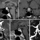
features, radiological profiles, pathological features and surgical outcomes of pituicytomas: a report of 11 cases and a pooled analysis of individual patient data. Radiological features of pituicytomas. a. The tumor is completely located in the intrasellar region. b. The tumor is located in the intrasellar-suprasellar region. c. The tumor is completely located in the suprasellar region. d. Pituicytoma invading the right cavernous sinus. The lesion surrounded the internal carotid artery, and the optic chiasm was not compressed. e. The optic chiasm was oppressed and elevated by the tumor, and the cavernous sinus was not involved. f. Pituicytoma invading the right cavernous sinus; the optic chiasm was compressed, and the tumor was accompanied by cystic degeneration

A rare case
of pituicytoma-related hypercortisolism in a patient with Cushing syndrome—case report. MRI T1-weighted sagittal view of non-enhancing 0.46 × 0.33 cm focus of low signal in the left central portion of the pituitary gland on the post-contrast

A rare case
of pituicytoma-related hypercortisolism in a patient with Cushing syndrome—case report. MRI T1-weighted coronal view same non-enhancing 0.46 × 0.33 cm pituitary lesion

A rare case
of pituicytoma-related hypercortisolism in a patient with Cushing syndrome—case report. MRI T1-weighted coronal view same non-enhancing 0.46 × 0.33 cm pituitary lesion and an arrow indicating the thickening of the pituitary stalk

Magnetic
resonance imaging of sellar and juxtasellar abnormalities in the paediatric population: an imaging review. Glioma in a 2-year-old girl with neurofibromatosis type 1. Sagittal (a) and axial (b) post contrast T1WI shows a partially enhancing lesion enlarging the optic chiasm and proximal optic nerves

Magnetic
resonance imaging of sellar and juxtasellar abnormalities in the paediatric population: an imaging review. Teratoma. Sagittal T1WI (a) and axial T2WI (b) show a lesion at the undersurface of the hypothalamus which has mixed low, intermediate and high signal. Axial CT image (c) shows the lesion to contain zones of calcification, fat and intermediate soft-tissue attenuation

Teenager with
short stature. Sagittal (above) and coronal (below) T1 MRI with contrast of the sella shows a large homogenous solid mass arising in the sella and extending superiorly into the suprasellar region that shows homogenous enhancement with contrast.The diagnosis was pituitary macroadenoma.

Magnetic
resonance imaging of sellar and juxtasellar abnormalities in the paediatric population: an imaging review. Germ cell tumours in two different children. Post-contrast T1WI (a) shows a large enhancing lesion in the suprasellar cistern extending superiorly into the third ventricle and posteriorly into the prepontine cistern. Sagittal post-contrast T1WI (b) in another child shows enhancing, disseminated germ-cell tumour in the suprasellar cistern, third and lateral ventricles, with involvement of the corpus callosum and optic chiasm

Magnetic
resonance imaging of sellar and juxtasellar abnormalities in the paediatric population: an imaging review. Enthesioneuroblastoma. Sagittal T1WI (a) and post-contrast T1WI (b) show a destructive mass lesion involving the sphenoid bone which invades the sphenoid sinus and sella. The tumour shows prominent, mildly heterogeneous contrast enhancement

Extraventricular
neurocytoma of the sellar region: case report and literature review. The pre- and post-operative MRI. a–d Preoperative MRI showing a intrasellar and suprasellar tumor extending to anterior skull base and superior clivus. e, f The postoperative enhanced MRI showing subtotal tumor removal, with a little tumor remained in the intrasellar region

Pituitary
region masses • Adamantinomatous craniopharyngioma - Ganzer Fall bei Radiopaedia

Pituitary
region masses • Pituitary macroadenoma - Ganzer Fall bei Radiopaedia

Pituitary
region masses • Suprasellar teratoma - Ganzer Fall bei Radiopaedia

Pituitary
region masses • Pituitary fossa meningioma - Ganzer Fall bei Radiopaedia

Pituitary
region masses • Rathke's cleft cyst - Ganzer Fall bei Radiopaedia

Pituitary
region masses • Suprasellar giant aneurysm - Ganzer Fall bei Radiopaedia

Pituitary
region mass (mnemonic) • Pituitary metastasis - Ganzer Fall bei Radiopaedia
Pituitary tumors, in other words, tumors which arise from the pituitary gland itself, include:
- pituitary adenoma
- pituitary carcinoma
- pituicytoma
- pituitary metastases
- spindle cell oncocytoma (rare)
- pituitary blastoma
Often the term is used more broadly to refer to pituitary region masses.
Siehe auch:
- Tuberkulose
- Meningeom
- Rathke Zyste
- Hirnmetastase
- Pilozytisches Astrozytom
- Raumforderungen des Clivus
- Aneurysma
- Chordom
- Hypophyse
- Makroadenom Hypophyse
- Teratom
- intrakranielle Lipome
- Chiasma opticum
- ektope Neurohypophyse
- verdickter Hypophysenstiel
- Pituitary MRI (an approach)
- Kraniopharyngeom
- Nervus opticus
- Germinom
- Sinus cavernosus
- dritter Ventrikel
- Mikroadenom Hypophyse
- Keimzelltumor
- Hypophysenadenom
- Erdheim-Chester-Erkrankung
- Opticusgliom
- hypothalamisches Hamartom
- hypothalamic lesions
- pituitary region mass with intrinsic high T1 signal
- intrakraniale Teratome
- skeletale Manifestationen der Langerhanszell-Histiozytose
- cystic meningioma
- solid and enhancing pituitary region mass
- Epidermoid
- normal bone marrow signal of the clivus
- intrakranielle Dermoidzyste
- Histiozytose
- Cisterna chiasmatica
- Granularzelltumor
- mixed cystic and solid pituitary region mass
- Pituizytom
- purely intrasellar pituitary mass
- zystische Läsionen der Sellaregion
- Spindelzell-Onkozytom der Hypophyse
- sakkuläre zerebrale Aneurysmata
- granular cell tumour of the pituitary
- Langerhans-Zell-Histiozytose der Hypophyse
- kongenitale Anomalien der Hypophyse
- supraselläres Teratom
- suprasellar lipoma
- Xanthogranulom der Hypophyse
- SATCHMO
- supraselläres Germinom
- cleft cyst
- hypothalamic astrocytoma / glioma
- meningioma, metastasis
- supraselläres Gangliogliom
- lymphozytäre Hypophysitis
- Metastasen in der Hypophyse
und weiter:
- Tumoren der Hypophysenregion
- Einklemmung
- Akromegalie
- suprasellar cistern lipoma
- Hypophysenstiel
- Hirntumoren
- zystische Läsionen in der Hypophyse
- intracranial choriocarcinoma
- Apoplex der Hypophyse
- mostly / purely cystic pituitary region masses
- transsphenoidale Hypophysektomie
- Galaktorrhö-Amenorrhö-Syndrom
- Chiasma-Syndrom
- suprasellar / hypothalamic lesions
- Pubertas praecox
- extra axial
- Hypophysitis
- teratoma suprasellar
- Hypophysenadenom Kalk
- Fröhlich-Syndrom
- Hypophysenkarzinom
- planum sphenoidale meningioma
- adamantinomatous craniopharyngioma
- cystic craniopharyngioma
- Germinom des ZNS
- kongenitale Anomalien der Sellaregion
- papillary craniopharyngioma
- intrakranieller Keimzelltumor
- Duplikation der Hypophyse
- Gangliogliom des Nervus opticus
- Hypophysentumor Estradiol
- Teratom der Hypophyse
- intracranial germ cell tumours
- Hirnherniation

 Assoziationen und Differentialdiagnosen zu Tumoren der Hypophysenregion:
skeletale
Manifestationen der Langerhanszell-Histiozytose


