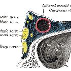
Hypophysenadenom


Pituitary adenomas are primary tumors that occur in the pituitary gland and are one of the most common intracranial neoplasms.
Depending on their size they are broadly classified into:
- pituitary microadenoma: less than 10 mm in size
- pituitary macroadenoma: greater than 10 mm in size
Although this distinction is largely arbitrary, it is commonly used and does highlight an important fact: small intrapituitary lesions (microadenomas) present differently and have different surgical and imaging challenges from larger lesions (macroadenomas) that extend into the suprasellar region. As such, it is not unreasonable to discuss them separately. This article is a general overview.
Epidemiology
Pituitary adenomas are common, with rates varying widely depending on the definition: population prevalence is approximately 0.1%; autopsy prevalence is around 15% . They account for approximately 10% of all intracranial neoplasms and 30-50% of all pituitary region masses .
Pituitary macroadenomas are approximately twice as common as microadenomas .
A minority of tumors are associated with multiple endocrine neoplasia type I (MEN I), multiple endocrine neoplasia type IV (MEN4), Carney complex, McCune-Albright syndrome, and familial isolated pituitary adenoma.
Clinical presentation
Pituitary adenomas present either due to hormonal imbalance (both microadenomas and macroadenomas) or mass effect on adjacent structures (macroadenomas), classically the optic chiasm. Rarely presentation can be catastrophic, due to pituitary apoplexy.
Hormone imbalance
Over half of all adenomas are secretory , although even when this is the case this may not be the cause of presentation. A lack of libido or even galactorrhea may not lead to presentation and as such many secreting tumors are only diagnosed when mass effect occurs (see below).
Hormones secreted include:
- secretory: ~65%
- prolactin: ~50%
- growth hormone (GH): 10%
- adrenocorticotropin (ACTH): 6%
- thyrotropin (TSH): 1%
- mixed
- non-secretory: ~35% most tend to be macroadenomas
It is also important to note that larger tumors can lead to hormonal imbalance due to mass effect rather than secretion. Hypopituitarism or moderately elevated prolactin are both seen, the later due to so-called stalk effect; prolactin release (unlike other pituitary hormones) is tonically inhibited by prolactin inhibitory hormone (PIH - a.k.a. dopamine) and as such compression of the pituitary infundibulum can result in elevation of systemic prolactin levels due to interruption of normal inhibition. It is also important to remember that numerous drugs that are dopamine antagonists will also elevate prolactin - see elevated prolactin (differential) .
Mass effect
Most of the cases presenting due to mass effect are due to non-secreting macroadenomas and the most common structure to be compressed by a macroadenoma is the optic chiasm. Invasion into the cavernous sinus is also encountered, with occasional compression of the oculomotor (CN III) or less frequently abducens (CN VI) nerves. Uncommonly large tumors may result in hydrocephalus (by compressing the midbrain or distorting the third ventricle), orbital or sinonasal symptoms.
Pathology
Location
Very rarely pituitary adenomas may be seen in extra-sellar ectopic locations, most commonly within sphenoid sinus. It may also be found in suprasellar region, cavernous sinus, parasellar region, clivus, nasal cavity, nasopharynx, temporal bone and third ventricle.
Radiographic features
Radiographic features are discussed separately:
Treatment and prognosis
Treatment of pituitary adenomas depends on a number of factors:
- size and presence of symptoms related to the mass effect: these will often necessitate surgical decompression regardless of cell type
- cell type: prolactin and growth hormone-secreting tumors can often be treated medically
Surgical management
The most commonly employed approach to pituitary masses is transsphenoidal, whereby the floor of the pituitary fossa is accessed via the nasal cavity. In large tumors, other approaches may be necessary (e.g. craniotomy).
Medical management
Medical management of prolactinomas relies on administering a dopamine receptor agonist (e.g. bromocriptine or cabergoline). Although it can dramatically reduce the size of a macroadenoma, it has been associated with an increased incidence of hemorrhage into the tumor .
Growth hormone-secreting tumors are usually surgically resected, however, in recurrent cases or in patients who are not able to undergo surgery they can be treated with octreotide (a long-acting somatostatin analog). This can result in both reduction of the size of the tumor and reduction in the serum levels of growth hormone .
Stereotactic radiosurgery
Radiosurgery is also occasionally used. Its main complications are hypopituitarism (seen in up to 70% of cases). Less common complications include damage to the optic apparatus (optic nerves, chiasm, optic tracts), cranial nerves and internal carotid arteries .
Prognosis
Recurrent symptoms requiring further intervention is relatively common, with 18% of patients with non-functioning tumors and 25% of patients with prolactinomas eventually needing further treatment .
Siehe auch:
- Hypophyse
- Tumoren der Hypophysenregion
- Makroadenom Hypophyse
- Teratom
- WHO-Klassifikation der Tumoren des zentralen Nervensystems
- Astrozytom
- Mikroadenom Hypophyse
- Verschlusshydrocephalus
- multiple endokrine Neoplasie Typ 1
- Nervus abducens
- Nervus oculomotorius
- Apoplex der Hypophyse
- papillärer Tumor der Hypophysenregion
- lymphozytäre Hypophysitis
und weiter:
- Morbus Basedow
- Tumor Kleinhirnbrückenwinkel
- neuroradiologisches Curriculum
- Gynäkomastie
- Makroorchidie
- Somatostatin-Rezeptor-Szintigrafie
- Pituizytom
- Carney-Komplex
- mass involving the foramen of Monro or/and superior third ventricle
- Hypophysitis
- Achard-Thiers-Syndrom
- Hypogonadotroper Hypogonadismus
- Hypophysenadenom Kalk
- Hypophysenkarzinom
- MRI protocol pituitary gland
- deviated pituitary stalk
- Hirsutismus
- zystisches Hypophysenadenom
- Lymphom der Hypophyse
- Cushing-Syndrom
- cavernous sinus invasion by a pituitary adenoma
- pituitary adenoma - necrotic
- Hypophysenadenom Kontrastmittel
- Metastasen in der Hypophyse

 Assoziationen und Differentialdiagnosen zu Hypophysenadenom:


