cavernous sinus

The cavernous sinuses are paired dural venous sinuses.
Gross anatomy
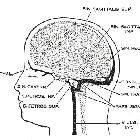
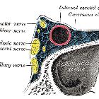
The cavernous sinus is located on either side of the pituitary fossa and body of the sphenoid bone between the endosteal and meningeal layers of the dura. It spans from the apex of the orbit to the apex of the petrous temporal bone. Unlike other dural venous sinuses, it is divided by numerous fibrous septa into a series of small caves, which is where its name is derived from. The normal lateral wall should be either straight or concave.
Boundaries
- roof: fold of dura mater attached to the anterior and middle clinoid processes
- anterior wall: medial end of the superior orbital fissure
- posterior wall: petrous apex
- medial wall: endosteum overlying the body of the sphenoid bone
- lateral wall: dura mater from the ridge of the roof to the floor of the middle cranial fossa
- floor: endosteum overlying the base of the greater wing of the sphenoid bone
Relations
- superiorly: middle cerebral artery, optic chiasm
- anteriorly: apex of the orbit
- posteriorly: cerebral peduncle
- medially: pituitary fossa, pituitary gland
- laterally: temporal lobe (medial surface), Meckel's cave (posteroinferiorly)
- inferiorly: sphenoid sinus
Vascular connections
It receives venous blood from:
- inferior and superior ophthalmic veins
- intercavernous sinus
- sphenoparietal sinus
- superficial middle cerebral vein
- occasionally
- central retinal vein
- a frontal tributary of the middle meningeal vein
Drainage of the cavernous sinus is via:
- superior petrosal sinus to the transverse sinus
- inferior petrosal sinus directly to the jugular bulb
- venous plexus on the internal carotid artery (ICA) to the clival (basilar) venous plexuses
- emissary veins passing through the
- foramen Vesalii
- foramen ovale: communicates between the CS and pterygoid venous plexus
- foramen lacerum
Depending on relative pressures the superior ophthalmic veins either drain to or from the cavernous sinus.
Additionally, the cavernous sinuses connect to each other via the intercavernous sinuses.
Contents
These can be remembered with the mnemonic O TOM CAT.
Nerves
The cavernous sinus transmits multiple cranial nerves to the superior orbital fissure and foramen rotundum. These are:
- in the lateral wall from superior to inferior
- oculomotor nerve (CN III)
- trochlear nerve (CN IV)
- trigeminal nerve (CN V)
- ophthalmic division
- maxillary division: within the very inferolateral aspect of the cavernous sinus wall or even outside the sinus rather than truly within it
- traversing the sinus
- abducens nerve (CN VI): inferolateral to the ICA
Artery
The ICA enters the posterior inferior aspect of the sinus and bends upon itself as the carotid siphon (cavernous segment - C4). Two branches arise from this segment: meningohypophyseal trunk and inferolateral trunk.
The artery is surrounded by a plexus of sympathetic nerves from the superior cervical ganglion.
Fat
Fatty deposits may be present within the cavernous sinus, especially in obese patients or in those who are taking corticosteroids .
Related pathology
Siehe auch:
- Arteria carotis interna
- Nervus trigeminus
- Sinus durae matris
- Sinus transversus
- Fissura orbitalis superior
- superficial middle cerebral vein
- meningohypophyseal trunk
- inferior petrosal sinus
- superior petrosal sinus
- Nervus oculomotorius
- Nervus abducens
- inferolateral trunk
- Raumforderungen im oder am Sinus cavernosus
- Sinus sphenoparietalis
- Nervus trochlearis
- Carotis-Sinus-cavernosus-Fistel
- Sinus-cavernosus-Syndrom
- Sinus cavernosus Thrombose
- dura
- OTOM CAT
und weiter:
- intrakranielle Verkalkungen
- Tumoren der Hypophysenregion
- durale AV-Fistel
- Brain stone
- Fossa pterygopalatina
- Cavum trigeminale
- basilar venous plexus
- pituitary MRI - an approach
- intercavernous sinus
- Vena ophthalmica superior
- cavernous sinus (mnemonic)
- cavernous sinus meningioma
- dilatierte Vena ophthalmica superior
- Raumforderung Sinus cavernosus
- cavernous sinus invasion by a pituitary adenoma


 Assoziationen und Differentialdiagnosen zu Sinus cavernosus: