third ventricle


The third ventricle is one of the four CSF-filled cavities that together comprise the ventricular system.
Gross anatomy
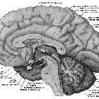
The third ventricle is a median cleft between the two thalami, which make up the superior aspect of the lateral walls. The thalami are separated from the hypothalamus (anteriorly) and subthalamus (posteriorly) by the hypothalamic sulcus.
The anterior wall of the third ventricle is formed from superior to inferior by the columns of the fornix, the anterior commissure and the lamina terminalis.
Posteriorly, it is bounded from superior to inferior by the habenular commissure, the pineal gland and the posterior commissure.
The roof is formed by the choroidal fissure and the bodies of the fornix.
The floor is bounded from anterior to posterior by the optic chiasm (posterior aspect), the infundibulum of the pituitary gland, the tuber cinereum, the mammillary bodies.
There are two small anterior recesses and two small posterior recesses:
- anterior:
- supra-optic recess (above the optic chiasm)
- infundibular recess (above the pituitary stalk)
- posterior:
The third ventricle contains choroid plexus along its roof along the tela choroidea continuous with the choroid plexus from the lateral ventricles.
It communicates with the:
- lateral ventricles via the foramina of Monro (interventricular foramina) lying just posterior to the columns of the fornix
- fourth ventricle via the aqueduct of Sylvius (cerebral aqueduct) just below the posterior commissure
Variant anatomy
In some patients, the two thalami meet forming the interthalamic adhesion and when casts are made of the ventricular system, a hole is seen through the third ventricle.
The infundibular recess can vary in its invagination into the infundibulum, most commonly extending the entire length, less commonly 50% or <50% of the length of the infundibular stalk.
Siehe auch:
- Thalamus
- Hypothalamus
- Seitenventrikel
- Hypophysenstiel
- vierter Ventrikel
- Kolloidzyste des dritten Ventrikels
- Hirnventrikel
- Recessus chiasmaticus
- Foramen interventriculare Monroi
und weiter:
- Tumoren der Hypophysenregion
- Pituitary MRI (an approach)
- Cisterna quadrigeminalis
- velum interpositum
- internal cerebral vein
- Verschlusshydrocephalus
- ambient cistern
- Hirntumoren
- medial posterior choroidal artery
- pituitary MRI - an approach
- third ventriculostomy
- aqueduct of Sylvius
- communicating obstructive hydrocephalus
- transcallosal approach
- Hydrocephalus communicans


 Assoziationen und Differentialdiagnosen zu dritter Ventrikel:


