optic nerve


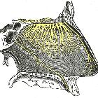
The optic nerve is the second cranial nerve which, along with the olfactory nerve (CN I), is really an extension of the central nervous system. It is not surrounded by Schwann cells with the first sensory bipolar cell body located peripherally in the retina. Their central processes synapse on ganglion cells on the vitreous surface of the retina and their central processes pass via the optic disc out of the globe and form the optic nerve proper.
The optic nerve is divided into four segments:
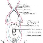
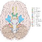
At the optic chiasm, the nasal fibers of each optic nerve (fibers carrying light impulses from the nasal side of the retina) decussate while the temporal fibers do not (partial decussation). From the optic chiasm arise two optic tracts, each one containing nasal fibers of the contralateral optic nerve and temporal fibers from the ipsilateral optic nerve. The optic tract courses around the cerebral peduncle to relay in the lateral geniculate body of the thalamus.
Arising from the lateral geniculate body of the thalamus, the optic radiations are divided into superior and inferior bundles. The superior bundle carries information from the superior retinal quadrant that represents the inferior visual field and ends at the superior aspect of calcarine sulcus (cuneus). The inferior bundle (including Meyer loop) carries information from the inferior retinal quadrant which represents the superior visual field. It passes anteriorly in the temporal lobe and forms the lateral wall of the inferior horn of lateral ventricle before passing posteriorly to end inferior to the calcarine sulcus (lingual gyrus).
Blood supply
Intraocular, intraorbital, and intracanalicular segments are supplied by the ophthalmic artery and its branch, the central retinal artery.
Small branches of the ACA and the superior hypophyseal artery supply the intracranial segment of the optic nerves and optic chiasm.
The optic tracts are supplied by small branches of the anterior choroidal and PCOM arteries.
Variant anatomy
According to a study by Delano et al., the course of the optic nerve in relation to the sphenoid sinus was classified according to four types :
- type 1: most common (76%): the optic nerve is immediately adjacent to the lateral or superior wall of the sphenoidal sinus, without impression on the sinus wall
- type 2: (15%): nerve causes an impression on the lateral sphenoidal sinus wall
- type 3: (6%): nerve courses through the sphenoidal sinus rather than simply running adjacent to the sinus
- type 4: (3%) nerve courses immediately lateral to the posterior ethmoidal and sphenoidal sinuses
Related pathology
Siehe auch:
- Hydrocephalus
- Dura mater
- Nervus olfactorius
- subarachnoid space
- Hirnnerven
- Opticusgliom
- Meningeom Nervus opticus
- Annulus of Zinn
- Orbita
- Papillenödem
- Sehbahn
- verdickter Nervus Opticus
- Nervus opticus Hämatom
- kraniofaziale fibröse Dysplasie Nervus opticus
und weiter:


 Assoziationen und Differentialdiagnosen zu Nervus opticus: