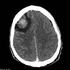
intrakranieller Keimzelltumor


Intracranial germ cell tumors are a heterogeneous group with variable imaging appearances, biology, response to treatment and prognosis.
The WHO classification of CNS tumors divides intracranial germ cell tumors into:
- germinoma (account for 60-80% of all cases)
- embryonal carcinoma
- yolk sac tumor
- choriocarcinoma
- teratoma
- immature
- mature
- teratoma with malignant transformation
- mixed germ cell
Epidemiology
Intracranial germ cell tumors make up approximately 0.4 to 1% of brain tumors in the western population, although the incidence is up to 8 times higher in the far east (as is the rate of testicular tumors) .
In general, males are more frequently affected than females, and this is most evident in non-germinomatous tumors of the pineal, where approximately 90% are found in males . Interestingly this is not the case in suprasellar tumors, which have a relatively even distribution.
Overall peak in incidence around the time of puberty (10-19 years of age), somewhat earlier for non-germinomatous germ cell and somewhat later for germinomas.
Clinical presentation
As is the case with other pineal region masses, intracranial germinomas tend to cause obstructive hydrocephalus due to compression of the aqueduct, and thus present with signs and symptoms of raised intracranial pressure.
Pathology
Markers
Tumor markers are useful in aiding preoperative diagnosis and monitoring following treatment. The two main markers are alpha-fetoprotein (AFP) which is synthesized by the yolk sac, and human chorionic gonadotropin (HCG) which is synthesized by the chorioepithelium .
- germinoma: usually neither AFP or HCG
- embryonal carcinoma: variable
- yolk sac tumor: AFP
- choriocarcinoma: HCG
- teratoma: variable, depending on tissues present
Radiographic features
These tumors tend to cluster in the midline, with a predilection of the pineal and the suprasellar regions. Controversy persists as to whether multifocal lesions, found at the time of diagnosis in 5-10% of patients, represents synchronous tumors or spread .
- pineal region: twice as common as all other sites
- suprasellar region
- next most common
- suprasellar germinomas more common in women
- floor of the third ventricle
- basal ganglia: more likely germinoma
- thalamus: more likely germinoma
- fourth ventricle
CT
CT is usually the first investigation that reveals an abnormality. Except mature teratomas, which often demonstrate fat density, CT cannot reliably differentiate between different types of germ cell tumors, although as a general rule germinomas are more homogeneous in appearance than non-germinoma germ cell tumors .
General features of intracranial germ cell tumors include:
- hyperdense compared to normal brain
- vivid contrast enhancement
- calcification: present in the majority of cases, usually representing engulfed normal pineal calcification, as well as sometimes tumor calcification .
Features of individual histologies are discussed separately.
MRI
MRI is the modality of choice for evaluation of pituitary region masses and pineal region masses. Similar to CT, it is difficult to distinguish histologies based on MRI appearance (again, except for the identification of fat in mature teratomas).
General imaging features include :
- T1: isointense to grey matter
- T2: isointense to grey matter
- T1 C+
- vivid contrast enhancement
- germinomas tend to be homogeneous
- DWI: restriction is common especially for germinomas due to high cellularity
- ADC values are higher than found in pineoblastoma
- SWI: hemorrhage is common in non-germinomatous germ cell tumors
Features of individual histologies are discussed separately.
Treatment and prognosis
In general, a biopsy is required as treatment and prognosis will vary with histology (whereas it is independent of location). Aggressive surgical debulking is of unproven benefit and carries significant morbidity given the usual locations of these tumors.
Germinomas are exquisitely radiosensitive, with cure achieved with radiation alone in 80-90% of patients .
Non-germinomatous tumors have much worse prognosis with survival rates ranging between 40-70% .
Teratoma prognosis depends on the degree of differentiation (mature vs. immature) and whether malignant transformation on a component is present (uncommon). In general, mature teratomas are indolent, whereas immature teratomas do poorly, with survival rates ranging between 50-70% .
Siehe auch:
- Tumoren der Hypophysenregion
- Teratom
- WHO-Klassifikation der Tumoren des zentralen Nervensystems
- Tumoren der Pinealisregion
- Germinom
- Chorionkarzinom
- Hodentumoren
- Hirntumoren
- basal ganglia germinoma
- hypothalamic germinoma
- Germinom des ZNS
- supraselläres Germinom
- Embryonales Karzinom
und weiter:

 Assoziationen und Differentialdiagnosen zu intrakranieller Keimzelltumor:


