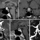
supraselläre Arachnoidalzyste

Suprasellar arachnoid cysts can be challenging to diagnose and, unlike many other arachnoid cysts, are usually symptomatic.
For a general discussion, please refer to the article on arachnoid cysts.
Clinical presentation
As can be expected from its location, suprasellar arachnoid cysts manifest clinically in the same way as other suprasellar masses, with visual symptoms due to compression and distortion of the optic pathways. Often these lesions extend superiorly, invaginating into the third ventricle and obstruct the outflow of the lateral ventricles, resulting in obstructive hydrocephalus .
Distortion of the pituitary infundibulum can also cause endocrine dysfunction .
Radiographic features
As with arachnoid cysts elsewhere, they are very thin walled (often imperceptible except on dedicated high-resolution T2 imaging) cystic lesions which follow CSF on all imaging modalities. There is no solid component and no enhancement (for further general discussion, please refer to the comprehensive article on arachnoid cysts).
These cysts invaginate superiorly into the third ventricle, and may even extend into one or both foramina of Monro. They are in such cases, associated with obstructive hydrocephalus.
Treatment and prognosis
Before the development of endoscopic neurosurgical techniques, treatment of these cysts could be challenging.
Current treatment of choice is to perform an endoscopic third ventriculostomy and creation of a communication between the third ventricle, cyst and basal cisterns, simultaneously treating the obstructive hydrocephalus and resolving the mass effect of the cyst itself .
Differential diagnosis
- obstructive hydrocephalus at the level of the aqueduct with the expansion of the third ventricle
- the floor of the third ventricle should be depressed
- no cyst wall should be visible or inferred (by CSF flow)
- cystic suprasellar lesions
- craniopharyngioma
- usually adamantinomatous subtype
- high protein content ("machinery oil")
- solid components
- calcification
- usually adamantinomatous subtype
- Rathke's cleft cyst
- the majority are completely or mostly intrasellar
- neurocysticercosis
- epidemiology features
- vesicular stage: cyst with dot sign
- craniopharyngioma
Siehe auch:

 Assoziationen und Differentialdiagnosen zu supraselläre Arachnoidalzyste:


