craniopharyngioma


Craniopharyngiomas are relatively benign (WHO grade I) neoplasms that typically arise in the sellar/suprasellar region. They account for ~1-5% of primary brain tumors, and can occur anywhere along the infundibulum (from the floor of the third ventricle to the pituitary gland).
There are two histological subtypes, adamantinomatous and papillary, which are said to differ not only in appearance but also in prognosis and epidemiology. A mixed or transitional subtype has also been described, although imaging features and prognosis are similar to the adamantinomatous subtype . Whether or not they represent distinct entities or a spectrum of morphology remains a bit controversial .
Epidemiology
The adamantinomatous subtype is more common than the papillary subtype by a 3-9-fold difference .
Although craniopharyngiomas are found in patients of all ages, there is a bimodal distribution . The first peak occurs between the ages of 5-15 years, consisting almost exclusively of the adamantinomatous subtype. A second smaller peak occurs in adults aged over 40 years old, consisting of both papillary and adamantinomatous subtypes . The papillary subtype is found almost exclusively in adults .
There is no gender predilection .
Clinical presentation
Clinical presentation is variable on account of the variable location and size of the tumor. Presenting complaints include:
- headaches and raised intracranial pressure
- visual symptoms
- 20% of children
- 80% of adults
- hormonal imbalances
- short stature and delayed puberty in children
- decreased libido
- amenorrhea
- diabetes insipidus
- behavioral change due to frontal or temporal extension
Pathology
Craniopharyngiomas are believed to derive from the Rathke cleft rather than squamous cell crests along the craniopharyngeal duct as was previously thought . Two distinct histological subtypes are recognized; adamantinomatous and papillary. Although they can sometimes coexist, these histological subtypes are usually fairly distinct with different demographics and imaging features.
Subtypes
Adamantinomatous
This type is seen predominantly in children. It consists of reticular epithelial cells that have appearances reminiscent of the enamel pulp of developing teeth.
There may be single or multiple cysts filled with thick oily fluid rich in protein, blood products, and/or cholesterol, giving the so-called "motor oil" fluid appearance. "Wet keratin nodules" are a characteristic histological feature. Calcification is usually present (~90%) .
Papillary
The papillary subtype is seen almost exclusively in adults and is formed of masses of metaplastic squamous cells . "Wet keratin" is absent. Cysts do form, but these are not a prominent feature, and the tumor is more solid. Calcification is uncommon or even rare .
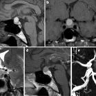
Radiographic features
Although similar in terms of location, radiographic features depend on the type, although due to a significant minority of tumors having both adamantinomatous and papillary components, they may show overlapping features.
Location
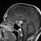
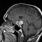
Craniopharyngiomas are primarily suprasellar tumors (75%) while a small intrasellar component is present in 20-25% of cases . Purely intrasellar location is quite uncommon (<5%), and may be associated with the expansion of the pituitary fossa . Larger tumors can extend in all directions, frequently distorting the optic chiasm or compressing the midbrain with resulting obstructive hydrocephalus.
Occasionally, craniopharyngiomas appear as intraventricular, homogeneous, soft-tissue masses without calcification (papillary subtype). The third ventricle is a particularly common location.
Rare or ectopic locations reported include nasopharynx, posterior fossa, extension down the cervical spine.
Adamantinomatous
Adamantinomatous craniopharyngiomas typically have a lobulated contour as a result of usually being multiple cystic lesions. Solid components are present, but often form a relatively minor part of the mass and enhance vividly on both CT and MRI. Overall, calcification is very common, but this is only true of the adamantinomatous subtype (~90% are calcified) .
These tumors have a predilection to being large, extending superiorly into the third ventricle, encasing vessels and even adhering to adjacent structures .
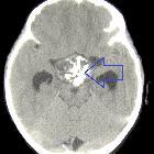
CT
- cysts
- near-CSF density
- typically large and a dominant feature
- present in 90% of cases
- solid component
- soft tissue density
- enhancement in 90%
- calcification
- seen in 90%
- typically stippled and often peripheral in location
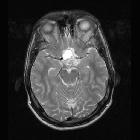
MRI
- cysts
- T1: iso- to hyperintense to brain (due to high protein content "motor oil cysts")
- T2: variable but ~80% are mostly or partly T2 hyperintense
- solid component
- T1 C+ (Gd): vivid enhancement
- T2: variable or mixed
- calcification
- difficult to appreciate on conventional imaging
- susceptible sequences may better demonstrate calcification
- MR angiography: may show displacement of the A1 segment of the anterior cerebral artery (ACA)
- MR spectroscopy: cyst contents may show a broad lipid spectrum, with an otherwise flat baseline
Papillary
Papillary craniopharyngiomas tend to be more spherical in outline and usually lack the prominent cystic component; most are either solid or contain a few smaller cysts. Calcification is uncommon or even rare in the papillary subtype, a fact has often forgotten .
These tumors tend to displace adjacent structures.
CT
- cysts
- small and not a significant feature
- near-CSF density
- solid component
- soft tissue density
- vivid enhancement
- calcification
- uncommon, rare
MRI
- cysts
- when present, they are variable in signal
- T1: 85% are T1 hypointense
- solid component
- T1: iso- to slightly hypointense to brain
- T1 C+ (Gd): vivid enhancement
- T2: variable/mixed
- MR spectroscopy: cyst contents does not show a broad lipid spectrum as they are filled with aqueous fluid
Treatment and prognosis
Treatment is usually surgical with radiotherapy, especially useful for incomplete resection. The surgical approach depends on the size and sellar vs suprasellar extent. Some lesions can be accessed via a transsphenoidal approach, whereas others require a craniotomy.
Benign local recurrence is seen in up to a third of patients and is said to significantly depend on histology: papillary has a much lower recurrence rate than adamantinomatous, although this may merely represent the more extensive and adherent character of the latter rather than an intrinsically more aggressive biological nature .
History and etymology
Craniopharyngiomas have also been known as pituitary ameloblastomas - there are no specific histological features to differentiate them from mandibular ameloblastomas .
Differential diagnosis
General imaging differential considerations include:
- Rathke cleft cyst
- no solid or enhancing component
- calcification is rare
- unilocular
- the majority are completely or mostly intrasellar
- pituitary macroadenoma (with cystic degeneration or necrosis)
- can look very similar
- usually has intrasellar epicenter with pituitary fossa enlargement rather than the suprasellar epicenter
- despite the occasional presence of T1 bright cystic regions, calcification in these cases is often absent (whereas most adamantinomatous craniopharyngiomas are calcified)
- intracranial teratoma
- presence of fat is helpful but requires fat-saturated sequences or CT to confirm
See also
- differential for a suprasellar mass
Siehe auch:
- Rathke Zyste
- Makroadenom Hypophyse
- Tumoren der Hypophysenregion
- intrakraniale Teratome
- adamantinomatous craniopharyngioma
- Xanthogranulom in der Sella
- papilläres Kraniopharyngeom
und weiter:
- Meningeom
- Raumforderungen des Clivus
- Hypophyse
- intraventrikuläre Neoplasien und Läsionen
- Empty-Sella-Syndrom
- ektope Neurohypophyse
- intrakranielle Epidermoidzyste
- WHO-Klassifikation der Tumoren des zentralen Nervensystems
- neuroradiologisches Curriculum
- Mikroadenom Hypophyse
- zystische Läsionen in der Hypophyse
- Rathke's pouch
- intraventrikuläre Neoplasien und Läsionen - Überblick
- pituitary region mass with intrinsic high T1 signal
- hypothalamic lesions
- intraventricular epidermoid
- solid and enhancing pituitary region mass
- beschleunigte Skelettreifung
- Apoplex der Hypophyse
- verzögerte Skelettreifung
- zystische Läsionen der Sellaregion
- purely intrasellar pituitary mass
- mixed cystic and solid pituitary region mass
- mostly / purely cystic pituitary region masses
- fetale Hirntumoren
- suprasellar / hypothalamic lesions
- mass involving the foramen of Monro or/and superior third ventricle
- Hypophysenadenom Kalk
- supraselläre Arachnoidalzyste
- cystic craniopharyngioma
- Lymphom der Hypophyse
- papillary craniopharyngioma
- MR spectroscopy and diffusion imaging of a craniopharyngioma
- Kalkkappe Kraniopharyngeom
- lymphozytäre Hypophysitis
- Metastasen in der Hypophyse


 Assoziationen und Differentialdiagnosen zu Kraniopharyngeom: