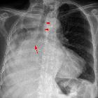
metastases to the pituitary gland


Pituitary metastases are rare, and unless a systemic metastatic disease is already apparent, are often preoperatively misdiagnosed as pituitary adenomas.
This article will discuss metastatic lesions affecting only the pituitary gland. For other intracranial metastatic locations, please refer to the main article on intracranial metastases.
Epidemiology
The demographics of affected patients reflect that of underlying primary tumors, which are most frequently breast cancer in women and lung cancer in men; thus elderly patients are most commonly affected . Pituitary metastases account for a minority of all intracranial metastases, although the figures vary widely from publication to publication .
Clinical presentation
Clinical presentation is variable but includes :
- hormonal dysfunction
- diabetes insipidus
- common: 29-71%
- presumably due to the predilection for posterior pituitary involvement (see below)
- rare symptom in pituitary adenomas, and can be a helpful distinguishing feature of pituitary metastasis
- panhypopituitarism
- hyperprolactinemia: disruption of the normal inhibition of prolactin release by dopamine
- diabetes insipidus
- mass effect
- optic chiasm compression
- extension into cavernous sinuses
Pathology
The most common primary malignancies metastasizing to the pituitary are breast cancer in women and lung cancer in men, presumably merely due to a large number of cerebral metastases from these two cancers . Many other primary tumors have also been described .
It is interesting to note that the posterior lobe and the infundibulum of the pituitary gland are more frequently involved than the anterior lobe (although this may not be the case in breast cancer ). Presumably due to the fact that the anterior pituitary receives its blood via the portal circulation rather than directly from the hypophyseal arteries .
Radiographic features
Although larger lesions are visible on CT, appearing as enhancing soft tissue masses, MRI is the modality of choice for assessment of the pituitary region.
MRI
Although all metastases to the pituitary (as is the case everywhere) start as microscopic deposits, they are usually encountered in two patterns:
Small intrasellar masses are generally not identified, mainly because they are presumably asymptomatic and require targeted sequences that are not performed without indication.
Sizable mass
These masses typically involve both the intra- and suprasellar compartments. As they are usually rapidly growing they have some features that are helpful in distinguishing them from pituitary macroadenomas:
- relatively normal size fossa (growth in a short period)
- bony destruction rather than remodeling
- dural thickening
- dumb-bell shape as the diaphragma sella has not had time to be stretched
- irregular edges
Infundibular lesion
Involvement of the infundibulum typically appears as nodular or irregular thickening and enhancement. The posterior pituitary bright spot may also be absent, either from interruption of the regular transport of neurosecretory granules down the infundibulum or due to concurrent infiltration of the posterior lobe.
Treatment and prognosis
Treatment is usually reserved for patients with symptomatic lesions (e.g. visual failure due to chiasmatic compression) or those in whom the diagnosis is not obvious (e.g. not known to have a malignancy, or thought to be in remission). Surgical decompression and biopsy in both cases can be carried out, although the overall prognosis and physical reserves of the patient need to be taken into account.
Whole brain radiotherapy is also an option when the pituitary lesion is one of many cerebral metastases . The proximity to the optic chiasm usually makes radiosurgery impractical without leading to loss of vision .
Prognosis is difficult to estimate as it will vary significantly depending on the systemic disease and the primary histology, although as a general ballpark figure a mean survival of approximately six months is in line with the published literature .
Differential diagnosis
The differential diagnosis of pituitary metastases is broadly that of pituitary region masses, and generally can be narrowed depending on the morphology of the lesion.
Solid and enhancing pituitary region masses have a differential diagnosis which includes:
- pituitary adenoma
- craniopharyngioma (papillary type)
- meningioma
- lymphocytic hypophysitis
- lymphoma
Nodular thickening and enhancement of the infundibulum have a differential diagnosis that includes :
- CNS tuberculosis
- Langerhans cell histiocytosis (eosinophilic granuloma)
- sarcoidosis
- lymphocytic hypophysitis
Siehe auch:
- Hirnmetastase
- Lungenkarzinom
- Sarkoidose
- Hypophyse
- Tumoren der Hypophysenregion
- verdickter Hypophysenstiel
- Tuberkulose des ZNS
- Kraniopharyngeom
- Hypophysenadenom
- Histiozytose X
- skeletale Manifestationen der Langerhanszell-Histiozytose
- Neoplasien der Mamma
- solid and enhancing pituitary region masses
- lymphozytäre Hypophysitis
und weiter:

 Assoziationen und Differentialdiagnosen zu Metastasen in der Hypophyse:


