intraventrikuläre Neoplasien und Läsionen - Überblick


intraventrikuläre Neoplasien und Läsionen - Überblick
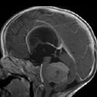
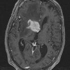
The ventricular system of the brain plays host to a variety of unique tumors, as well as tumors that are more frequently seen elsewhere (e.g. meningiomas). Besides, some intra-axial (parenchymal) masses can be mostly exophytic and thus appear mostly intraventricular. A systematic approach taking into account location, patient demographic and imaging appearances can often substantially narrow the differential, and in most cases suggest one diagnosis as by far the most likely. This is especially important if the mass is benign and can be safely ignored/observed.
Intraventricular masses
The main lesions to be considered are:
- tumors
- cysts
- colloid cyst
- intraventricular simple cysts (including arachnoid cysts, ependymal cysts and large choroid plexus cysts)
- choroid plexus xanthogranuloma
- cavum septum pellucidum
- cavum vergae
- cavum velum interpositum
- infection
- intraventricular hemorrhage
In addition, parenchymal lesions which can be periventricular should also be considered, such as:
- glioblastoma
- primary CNS lymphoma
- cerebral metastases
- medulloblastoma / sPNET
- hemangioblastoma
- pilocytic astrocytoma
- atypical teratoid / rhabdoid tumor
- pineal region masses
Imaging
As is the case with most intracranial pathology, MRI is the modality of choice for assessment of intraventricular masses, although CT and DSA both have roles to play. Transcranial ultrasound is particularly useful in infants.
A typical MRI protocol would include three plane imaging (essential if the relationship of the mass to the ventricle is to be confidently determined) and post-contrast studies (the pattern of enhancement is particularly useful in distinguishing some the lesions mentioned above).
Approach
Aunt Minnie lesions
Perhaps more so than in most other regions of the brain, many intraventricular masses have very characteristic appearances and offer little in the way of a realistic differential diagnosis (or at most between two lesions that are difficult to distinguish on imaging). These can be considered Aunt Minnies and the only way to approach them is to be familiar with their appearance. Examples of such lesions include:
- colloid cyst
- intraventricular simple cysts
- choroid plexus xanthogranuloma
- cavum septum pellucidum
- cavum vergae
- cavum velum interpositum
- subependymal giant cell astrocytoma / subependymal hamartomas of tuberous sclerosis
- central neurocytoma
Intraventricular vividly enhancing mass
Finding a vividly enhancing mass in the ventricular system has a limited differential, including:
- choroid plexus papilloma
- peak incidence: young children
- location: typically in the trigone of children and fourth ventricle of adults
- associated with hydrocephalus
- highly lobulated
- extremely vividly enhancing
- choroid plexus carcinoma can appear identical although usually there is evidence of heterogeneity (necrosis / hemorrhage) and brain invasion
- intraventricular meningioma
- peak incidence: middle age to older adults
- location: 85% in trigone of the lateral ventricle
- solid and well circumscribed and rounded / spherical or with a few large lobulations
- homogeneous signal intensity
- moderate restricted diffusion
- dense calcification is characteristic
- ependymoma
- peak incidence: children and young adults
- location: typically on the floor of 4 ventricle in children
- enhancement heterogeneous
- hemorrhage common
- metastasis
- peak incidence: usually older patients
- location: choroid plexus or anywhere with the ventricles
- heterogeneous
- often multiple lesions
- hemangioblastoma (not intraventricular, but can mimic an intraventricular mass)
- peak incidence: young adults
- location: posterior fossa
- cystic component
- large flow voids
Siehe auch:
- Xanthogranulome des Plexus choroideus
- Hirnmetastase
- Pilozytisches Astrozytom
- Glioblastoma multiforme
- Tuberöse Sklerose
- Ependymom
- Neurozystizerkose
- Oligodendrogliom
- intraventrikuläre Neoplasien und Läsionen
- Cavum septi pellucidi et vergae
- intraventrikuläre Blutung
- Medulloblastom
- Kraniopharyngeom
- Tumoren der Pinealisregion
- Hämangioblastom
- Cavum septi pellucidi
- primäres ZNS-Lymphom
- tuberculoma
- Karzinom des Plexus choroideus
- Plexuspapillom
- Kolloidzyste
- intraventrikuläres Meningeom
- Kolloidzyste des dritten Ventrikels
- intraventrikuläres Neurozytom
- Subependymom
- subependymales Riesenzellastrozytom
- intraventrikuläre Arachnoidalzyste
- intraventrikuläre Kolloidzyste
- Tumoren des Plexus choroideus
- Primitiver neuroektodermaler Tumor des ZNS
- atypischer teratoider rhabdoider Tumor
- Cavum veli interpositi Zyste
- Metastasen des Plexus choroideus
- Trigonum Syndrom (Seitenventrikel)
- Aunt Minnie
- primitive neuroectodermal tumour
und weiter:

 Assoziationen und Differentialdiagnosen zu intraventrikuläre Neoplasien und Läsionen - Überblick:


