Hemangioblastoma (central nervous system)


Hemangioblastomas are tumors of vascular origin and occur both sporadically and in patients with von Hippel Lindau (vHL). They are WHO grade I tumors that can occur in the central nervous system or elsewhere in the body, including kidneys, liver, and pancreas.
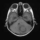
These tumors generally present on imaging as sharply demarcated homogeneous masses composed of a cyst with non-enhancing walls, a mural nodule which vividly enhances, often with prominent serpentine flow voids.
Epidemiology
Hemangioblastomas typically occur in young to middle-aged adults and, although they are the most common primary intra-axial infratentorial tumor in this demographic, they are nonetheless uncommon in absolute terms. They are estimated to account for only 1-2.5% of all intracranial tumors and approximately 10% of all posterior fossa tumors :
- slight male predilection in adults: M: F ratio of 1.3-2.6:1
- peak incidence at around 30-60 years of age, but earlier in patients with vHL
- sporadic cases make up approximately 75-80%, with the remainder being found in patients with vHL
Clinical presentation
Most frequently present with symptoms relating to:
- headaches: 70%
- hydrocephalus and symptoms of raised intracranial pressure: 50 %
- cerebellar dysfunction: ~50-60%
- altered mental state: 10%
- polycythemia due to erythropoietin production occurs in ~20% (range 5-40%) of cases
One study showed that in vHL patients, the onset of symptoms usually coincides with the development of a cystic component, and they showed that solid cerebellar nodules were mostly well-tolerated. They suggested repeat imaging at this stage to document the size of the cyst and to plan tumor excision .
Acute presentation due to significant hemorrhage is uncommon and tends to be seen in larger tumors (>1.5 cm) .
Pathology
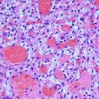
Hemangioblastoma is actually a capillary hemangioma and, despite the name with the affix of "blastoma", it is a low grade (WHO grade I) lesion (note that the calvarial hemangioma is a cavernous hemangioma). The tumor is usually well-circumscribed with a highly vascular mural nodule almost always abutting pial layer and a peripheral cyst which has similar contents as blood plasma. Hence it is suggested that the cystic component most likely arises by exudation from the solid nodule vascular component .
It has therefore also been suggested that resection of the cystic component need not be carried out. This has not been supported in a number of other studies however (a similar discussion is present in the pilocytic astrocytoma literature).
Cysts which are within the solid nodule, or have peripheral enhancement, are presumably part of the tumor and should, therefore, be resected. The fluid within the cyst is often xanthochromic due to frequent bleeding into the cyst .
Microscopy reveals a very vascular tumor composed of thin-walled vessels with surrounding stroma of connective tissue and polygonal lipid-laden 'stromal cells' .
Location
- intracranial: 87-97%
- 95% in the posterior fossa
- 85% in cerebellar hemisphere
- 10% in the cerebellar vermis
- 5% medulla
- only rarely do they extend beyond the cerebellum into the cerebellopontine angle
- 5% supratentorial (typically in the optic radiations)
- cerebral hemangioblastomas are only really seen in patients with vHL
- 95% in the posterior fossa
- spinal: 3-13%
Associations
- pheochromocytoma
- multiple RCCs
- von Hippel Lindau (vHL) disease
- ~45% of those with vHL develop hemangioblastomas
- ~20% of those with hemangioblastoma have vHL
- polycythemia: due to secretion of erythropoietin from lesions
Radiographic features
Typically hemangioblastomas (60% of cases) are sharply demarcated homogeneous masses composed of a cyst with non-enhancing walls, except for a mural nodule which vividly enhances and often has prominent serpentine flow voids . Not infrequently the mural nodule itself has cystic spaces within it. The solid nodules are commonly seen abutting the pia mater.
In the remaining 40%, the tumor is solid with no cystic cavity .
CT
- the mural nodule is isodense to the brain on non-contrast scans with fluid density surrounding cyst
- postcontrast intense homogeneous enhancement of the mural nodule
- the cyst walls do not usually enhance
- calcification is not a feature
MRI
- T1
- hypointense to isointense mural nodule
- CSF signal cyst content
- T1 C+ (Gd)
- mural nodule vividly enhances
- cyst wall does not enhance
- T2
- hyperintense mural nodule
- flow voids due to enlarged vessels may be evident especially at the periphery of the cyst, seen in 60-70% of cases
- fluid-filled cyst, similar to CSF
- MR perfusion imaging: high rCBV ratios
Angiography (DSA)
Enlarged feeding arteries and often dilated draining veins are demonstrated, with a dense tumor blush centrally .
Treatment and prognosis
Surgical resection is usually curative, and with large lesions may be made easier by preoperative embolization. Simple cyst drainage appears to be insufficient for definitive management. Adjuvant radiotherapy may be used in patients with incomplete resections. Recurrence can be seen in up to 25% of patients .
Differential diagnosis
General imaging differential considerations include:
- brain metastases: although single posterior fossa metastases are uncommon they are still the most common diagnosis if the patient is middle-aged or older
- astrocytoma
- pilocytic astrocytoma in children
- glioblastoma (GBM) in adults
- ependymoma
- vascular lesions
- arterio-venous malformation (AVM) with subacute bleed
- cavernoma with subacute bleed
- subacute infarction
- medulloblastoma
- adult patients
- much more solid
- restricted diffusion (small round blue cell tumor)
Siehe auch:
- Pilozytisches Astrozytom
- Glioblastoma multiforme
- Arteriovenöse Malformation
- Kavernom
- Ependymom
- Medulloblastom
- zystische Raumforderung hintere Schädelgrube
- Kleinhirntumoren
- spinales Hämangioblastom
- Tumoren der hinteren Schädelgrube
- infratentorielles Meningeom
- zystische zerebelläre Raumforderungen
- retinales Hämangioblastom
und weiter:
- Arachnoidalzyste
- vertebrale Metastasen
- intraventrikuläre Neoplasien und Läsionen
- Tumor Kleinhirnbrückenwinkel
- WHO-Klassifikation der Tumoren des zentralen Nervensystems
- Plexuspapillom
- intramedullary spinal metastasis
- Morbus Hippel-Lindau
- intraventrikuläre Neoplasien und Läsionen - Überblick
- meningeales Melanozytom
- spinale Metastasen
- cap sign of spinal tumour
- intradural spinal mass lesions - an approach
- spinales Pilozytisches Astrozytom
- Neoplasien der Cauda equina
- cystic haemangioblastoma of the medulla oblongata in Von Hippel-Lindau syndrome
- haemosiderin cap sign
- hemangioblastoma: von Hippel-Lindau syndrome
- Hämangioblastom der Medulla oblongata
- tumours associated with increased erythropoetin


 Assoziationen und Differentialdiagnosen zu Hämangioblastom: