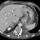
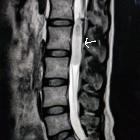
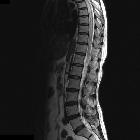
spinales Hämangioblastom


Spinal hemangioblastomas are the third most common intramedullary spinal neoplasm, representing 2-6% of all intramedullary tumors .
This article specifically relates to spinal hemangioblastomas. For a discussion on intracranial hemangioblastomas and a general discussion of the pathology refer to the main article: hemangioblastoma.
Epidemiology
Hemangioblastomas are mostly sporadic (two-thirds) with a peak presentation in the fourth decade. Sporadic hemangioblastomas only rarely occur in children. Males and females are affected equally .
One-third of patients with hemangioblastomas (not just spinal) have von Hippel-Lindau syndrome and typically these patients present earlier with multiple tumors.
Clinical presentation
Clinical presentation is similar to that of other intramedullary spinal tumors, with pain, weakness and sensory changes common. Rarely, spinal hemangioblastomas may cause subarachnoid hemorrhage or hematomyelia.
Pathology
Hemangioblastomas are benign vascular lesions and they do not undergo malignant degeneration, considered WHO grade I under the current (2016) WHO classification of CNS tumors .
Histologically, they consist of large pale stromal cells packed between blood vessels.
Radiographic features
The most common location is the thoracic cord (50%), followed by the cervical cord (40%) . The majority of hemangioblastomas have an intramedullary component with two-thirds located eccentrically and having an exophytic component (most commonly along the dorsum of the cord ). Only 25% are entirely intramedullary. A minority appears entirely extramedullary and only rarely are they extradural .
Eighty percent of hemangioblastomas are solitary. If multiple lesions are present, von Hippel-Lindau syndrome should be suspected .
Angiography
A densely enhancing nidus with associated dilated arteries and prominent draining veins is characteristic of a hemangioblastoma.
CT
On non-contrast CT they may be seen as a soft tissue nodule often with a prominent hypodense cyst-like component. Contrast administration results in vivid enhancement of the solid component.
MRI
Although they usually appear as discrete nodules, there can be diffuse cord expansion. An associated tumor cyst or syrinx is common (50-100%). Additionally, they may rarely be a source of subarachnoid hemorrhage or hematomyelia .
Reported signal characteristics of the solid components include :
- T1
- variable relative to the normal spinal cord
- hypo- to isointense most common, and difficult to identify
- hyperintense (25%)
- T2
- iso- to hyperintense
- focal flow voids especially in larger lesions
- surrounding edema and associated syrinx are usually seen
- hemosiderin capping may be present
- T1 C+ (Gd)
- the tumor nodule enhances vividly
Care should be taken to image the whole neuraxis to ensure that no other lesions are present.
Treatment and prognosis
Hemangioblastomas are slow-growing. They are usually treated by surgical resection, sometimes with preceding endovascular embolization to reduce intraoperative blood loss.
Differential diagnosis
The differential diagnosis can be thought of in two groups on account of the principal features of these tumors: neoplasms of the spinal canal (enhancing component) and vascular malformations of the spinal cord (enlarged vessels)
Other enhancing masses to be considered include:
Vascular malformation to consider include:
- other hypervascular cord neoplasms
- spinal arteriovenous malformation
- spinal dural arteriovenous fistula
- spinal cavernous malformations
Siehe auch:
- Hämangioblastom
- spinale Schwannome
- Ventriculus terminalis
- spinal paraganglioma
- neoplasms of the spinal canal
- Rückenmarksinfarkt
- spinal neurofibroma
- spinales Ependymom
- intraspinales Meningeom
- intramedulläre spinale Tumoren
- spinale durale arteriovenöse Fistel
- spinales Ependymom des Filum terminale
- spinale kavernomatöse Malformationen
- spinale Meningeosis neoplastica
- spinale arteriovenöse Malformationen
- spinales Astrozytom
- intramedullary metastases (spinal)
- intradurale extramedulläre Tumoren
und weiter:

 Assoziationen und Differentialdiagnosen zu spinales Hämangioblastom:


