intramedulläre spinale Tumoren

Radiological
approach to non-compressive myelopathies. Intramedullary ependymoma in a 6-year-old girl. Sagittal T2W (A) and T1W (B) images demonstrate a well-marginated oval-shaped lesion in the dorsal spinal cord (white arrow) extending from D8–D9 vertebral levels with associated cord expansion and perilesional edema. The lesion appears hyperintense on the T2W image with hypointense areas in the center (asterisk) which also shows a hyperintense signal on the T1W (B) image likely due to intratumoral hemorrhage. A T2 hypointense rim noted at superior and inferior poles of the lesion represents the hemosiderin cap (yellow arrow). Sagittal T1 fat saturated post-contrast image (C) demonstrates mild peripheral enhancement in the lesion (arrow)

Location,
length, and enhancement: systematic approach to differentiating intramedullary spinal cord lesions. Glioblastoma multiforme. A 46-year-old female with lumbar back pain and recent onset of urinary retention. There is an expansile lesion within the distal spinal cord with intermediate T2 hyperintensity (a, arrow) and non-uniform enhancement (c, arrowhead)

Intramedullary
spinal tumors • Cord compression from hemangioblastoma - Ganzer Fall bei Radiopaedia

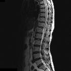
Primary
spinal cord oligodendroglioma: a case report and review of the literature. An MRI of the cervicothoracic spinal cord. Gadolinium-enhanced sagittal T1-weighted image (a) showed an enhanced intramedullary tumor from C2 to T4 level. Sagittal T2-weighted image (b) showed syringomyelia above tumor

Spinal
ependymoma • Ependymoma (cervical cord) - Ganzer Fall bei Radiopaedia

Ganglioglioma
• Ganglioglioma - cervical cord - Ganzer Fall bei Radiopaedia

Intramedullary
spinal tumors • Spinal pilocytic astrocytoma - Ganzer Fall bei Radiopaedia
intramedulläre spinale Tumoren
Siehe auch:
- Kavernom
- durale AV-Fistel
- Transverse Myelitis
- Encephalomyelitis disseminata
- neoplasms of the spinal canal
- spinales Hämangioblastom
- spinale Epidermoidzyste
- spinale durale arteriovenöse Fistel
- spinale Dermoidzyste
- spinales Astrozytom
- intramedullary metastases (spinal)
- intraspinale Tumoren
- intradurale extramedulläre Tumoren
- intramedulläres Ependymom
- intramedulläres Astrozytom
- Sarkoidose Rückenmark
- intramedulläre spinale Tuberkulose
- spinales Oligodendrogliom
und weiter:


 Assoziationen und Differentialdiagnosen zu intramedulläre spinale Tumoren:
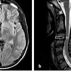


intramedullary
metastases (spinal)
