spinale durale arteriovenöse Fistel


Spinal dural arteriovenous fistulas (SDAVF) are the most common type of spinal vascular malformation, accounting for ~70% of all such lesions.
This article specifically relates to spinal dural arteriovenous fistulas. For a discussion of intracranial dural arteriovenous fistulas refer to the main article: dural arteriovenous fistula.
Epidemiology
The incidence of SDAVF peaks in the 5 and 6 decades, and males are more commonly affected than females .
Clinical presentation
SDAVFs cause symptoms through venous hypertension and congestion of the cord with edema. The most common clinical presentations are progressive pain, lower extremity weakness or sensory changes. Sphincter dysfunction may also occur. The onset of symptoms is insidious and progression occurs over several years. There is often a significant delay between presentation and diagnosis.
Hemorrhage is very rare but occasionally encountered, and may be a source of unexplained subarachnoid hemorrhage .
Pathology
These fistulae are abnormal direct connections between a radicular/radiculomeningeal artery and a radicular/pial vein in the dura of an adjacent nerve root sleeve. 85% of SDAVFs consist of a single transdural arterial feeder, however, there are cases with many arterial feeders originating from either a single or multiple levels that may be either unilateral or bilateral .
The direct arterial inflow into the venous system raises the pressure within the coronal venous plexus, which is valveless. The coronal venous plexus dilates and venous drainage of the cord decreases, causing venous congestion and intramedullary edema (congestive or venous hypertensive myelopathy). Cord ischemia and infarction may result. Nearly 60% of SDAVFs are spontaneous, with the remainder being caused by trauma .
Radiographic features
MRI
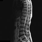
There is typically cord enlargement in the lower thoracic region and conus medullaris, with signal change involving multiple spinal segments. The segmental level of cord enlargement and signal change does not correlate with the location of the fistula .
Signal characteristics
- T1: intramedullary hypointensity and flow voids on the cord surface may be seen
- T2
- diffuse multilevel intramedullary hyperintensity (edema) - most sensitive finding . Regardless of the location of the fistula, the T2 hyperintensity involves the conus medullaris in up to 90% of cases because of orthostasis. The exception is SDAVFs of the upper cervical spine (C1-C2) which often drain intracranially and present more commonly with subarachnoid hemorrhage
- T2 hypointensity in the periphery of the cord . This is thought to represent pial capillaries containing deoxyhemoglobin secondary to venous hypertension
- prominent serpiginous intradural extramedullary flow voids - most specific finding. These usually span more than three segments on the dorsal and sometimes ventral aspects of the cord. The vessels may be large enough to give the surface of the cord a scalloped appearance. If there is significant cord swelling, the veins can be compressed by the mass effect and not be detectable on imaging .
- T1 C+ (Gd)
- patchy intramedullary enhancement is often seen (due to the breakdown of the blood-brain barrier because of either chronic infarction or a capillary leak phenomenon secondary to venous hypertension)
- serpentine enhancing veins on the cord surface
- contrast-enhanced MRA
- helps determine the segmental level of the fistula in order to guide selective catheter angiography
If MRI is contraindicated, the following may be performed:
- CT angiography: may successfully localize the fistula in up to 75% of cases
- CT myelography: may demonstrate tortuous filling defects due to dilated veins
Angiography (DSA)
DSA is the gold standard test for confirming the diagnosis and it provides with options for treatment. It is a time consuming and potentially dangerous investigation as dissection of a vessel can potentially lead to cord ischemia.
The site of maximal MRI abnormality is not a reliable indicator of the location of the fistula, which can be many levels away. As such a complete spinal angiogram consists of selective catheterization of the bilateral :
- intercostal arteries
- lumbar arteries
- median and lateral sacral arteries
- vertebral arteries
- ascending cervical arteries
- intracranial vessels may also need to be assessed if no fistula is found including
- ascending pharyngeal artery
- meningohypophyseal trunk
- middle meningeal artery
- occipital artery
Treatment and prognosis
Treatment options include endovascular or surgical occlusion of the shunt.
- endovascular occlusion
- performed with either cyanoacrylate glue or Onyx after superselective catheterization of the radiculomeningeal artery supplying the fistula
- occlusion rates of up to 85% have been reported
- endovascular treatment is contraindicated if the radicular artery also supplies the anterior spinal artery; embolization of a fistula that supplies a posterior spinal artery remains controversial
- surgery: surgical occlusion consists of a targeted laminectomy and intradural exploration with coagulation or disconnection of the draining vein; occlusion rates as high as 98% have been reported
After treatment of the fistula, the T2 hyperintensity, prominent flow voids, and enhancement should decrease with time but can persist for up to a year. These postoperative imaging features do not correlate with clinical outcome .
If treated early, motor and sensory function can be improved or stabilized in most cases. Pain and bowel and bladder dysfunction are only reversed in a minority of patients .
Differential diagnosis
General imaging differential considerations include:
- intramedullary neoplasm
- CSF flow artifact
- typically seen in the subarachnoid space dorsal to the cord
- no signal abnormality within the cord
- spinal arteriovenous malformation (AVM)
- hemorrhage is common
- acute onset of symptoms
- males and females affected equally
- usually presents during the 3decade
Siehe auch:
- Kaudaredundanz
- durale AV-Fistel
- Cognard classification of dural arteriovenous fistulas
- intramedulläre spinale Tumoren
- Borden classification of dural arteriovenous fistulas
- spinale arteriovenöse Malformationen
- kraniale durale AV-Fistel
und weiter:


 Assoziationen und Differentialdiagnosen zu spinale durale arteriovenöse Fistel:


