intradurale extramedulläre Tumoren

Cervical
intradural meningioma presenting as Brown-Séquard syndrome. Axial T1 after gadolinium injection. The mass is homogeneously enhancing and completely intradural, without extension to the foramen.

Cervical
intradural meningioma presenting as Brown-Séquard syndrome. Axial T1 at the level of C2, showing an intradural extramedullary mass occupying most of the canal and displacing the spinal cord posteriorly and to the right.

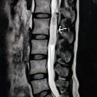
Cervical
intradural meningioma presenting as Brown-Séquard syndrome. Midline sagittal T1, showing an intradural extramedullary anterior mass occupying most of the canal and displacing the spinal cord posteriorly.

Cervical
intradural meningioma presenting as Brown-Séquard syndrome. Midline sagittal T2WI. Anterior intradural extramedullary lesion. Notice a line of CSF separating the lesion from the cord, displaced posteriorly.

Cervical
intradural meningioma presenting as Brown-Séquard syndrome. Axial T2WI at the level of C2, showing the intradural extramedullary mass anteriorly, displacing the cord posteriorly and to the right. It has a slight hyperintense signal intensity when compared to the cord parenchyma.

Cervical
intradural meningioma presenting as Brown-Séquard syndrome. Midsagittal T1WI. Homogeneous enhancement after iv gadolinium administration. Notice the dural tail in the anterior aspect of the dura, adjacent to the mass. There is no cord enhancement.

Cervical
intradural meningioma presenting as Brown-Séquard syndrome. Coronal T1WI. Homogeneous enhancement after iv gadolinium administration. The mass has well defined margins and does not invade the bone.
intradurale extramedulläre Tumoren
Siehe auch:
- Meningeom
- spinale Schwannome
- spinale Arachnoidalzyste
- spinal paraganglioma
- Dural-Tail-Zeichen
- neoplasms of the spinal canal
- spinal neurofibroma
- spinale Epidermoidzyste
- intradural extramedullary metastases
- intraspinales Meningeom
- intramedulläre spinale Tumoren
- spinales Ependymom des Filum terminale
- spinale Dermoidzyste
- spinal intradural extramedullary haemangiomas
- intradurales spinales Lipom
- dumbbell appearance
und weiter:


 Assoziationen und Differentialdiagnosen zu intradurale extramedulläre Tumoren: