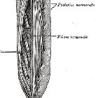
Ventriculus terminalis


The ventriculus terminalis (or persistent terminal ventricle, or terminal ventricle of Krause, or 5ventricle) is an ependymal-lined fusiform dilatation of the terminal central canal of the spinal cord, positioned at the transition from the tip of the conus medullaris to the origin of the filum terminale. This differs from a filar cyst which is located within the filum terminale.
It represents the canalization and retrogressive differentiation of the caudal end of the developing spinal cord and regresses in size during the first weeks after birth.
Radiographic features
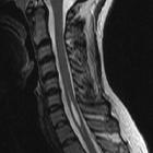
Irrespective of the modality used to image the spine, a ventriculus terminalis in newborns appears as a cystic structure at the tip of the conus medullaris, extending over 8-10 mm with a transverse diameter of 2-4 mm. Later in childhood it often remains visible as a tiny cystic structure but is rarely identifiable in adults.
MRI
Typically show fluid signal characteristics
- T1
- typically hypointense
- T2
- typically hyperintense
- T1 C+ (Gd): generally do not enhance
History and etymology
In 1859 the German anatomist Benedikt Stilling (1810-1879) , wrote that the ventriculus terminalis was an expanded CSF-space in the distal spinal cord with an ependymal lining; interestingly at that time it was called the seventh ventricle! Another German anatomist, Wilhelm Krause, determined that this was a true ventricle and named it the fifth ventricle .
Related pathology
Abnormal persistence of cystic dilatation of the ventriculus terminalis (cyst of the medullary conus) can occur and may present symptomatically in adulthood with bladder or bowel sphincter disturbance.
Siehe auch:
- Cavum septi pellucidi et vergae
- Syrinx
- Conus medullaris
- Syringomyelie
- Rückenmarksinfarkt
- spinales Hämangioblastom
- spinale Epidermoidzyste
- intraspinale zystische Läsionen
- Entwicklungsstörungen des Ventrikelsystems
und weiter:


 Assoziationen und Differentialdiagnosen zu Ventriculus terminalis: