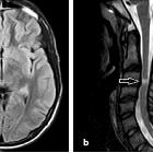
neurosarcoidosis


Central nervous system involvement by sarcoidosis, also termed neurosarcoidosis, is relatively common among patients with systemic sarcoidosis and has a bewildering variety of manifestations, often making diagnosis difficult.
For a general discussion of the underlying condition, please refer to the article sarcoidosis.
Epidemiology
The demographics of affected patients is similar to that of systemic sarcoidosis, typically affecting patients 30-40 years of age with a female predilection .
Clinical presentation
Central nervous system involvement by sarcoidosis is very variable, with lesions potentially involving the leptomeninges, pituitary and parenchyma of all parts of the intracranial compartment. Thus, clinical presentation is also very variable and nonspecific:
- signs and symptoms of raised intracranial pressure due to hydrocephalus
- cranial nerve palsies
- optic nerve involvement (particularly common)
- facial nerve palsy
- endocrine features of hypothalamic/pituitary sarcoidosis
- seizures
- variable weakness, paresthesias and dysarthria/dysphagia
- spinal cord involvement presenting as myelopathy
Although it is very rare (range 1-17% ) to have isolated neurosarcoidosis (i.e. without systemic disease), central nervous system symptoms are not uncommonly the first manifestation, and as such patients are often imaged without the diagnosis of systemic sarcoidosis having yet been made.
Interestingly up to 10% of patients with the systemic disease will demonstrate positive imaging findings; thus not all patients with demonstrable imaging findings of neurosarcoidosis are symptomatic.
Pathology
Histologically, central nervous system involvement is seen in ~20% (range 14-27%) of patients with systemic sarcoidosis, although only ~10% (range 3-15%) are symptomatic .
Radiographic features
The radiographic features of neurosarcoidosis can be thought of as occurring in one or more of five compartments. From superficial to deep they are:
- skull vault involvement (refer to musculoskeletal manifestations of sarcoidosis)
- pachymeningeal involvement
- leptomeningeal involvement (seen in up to 40% of cases )
- pituitary and hypothalamic involvement
- cranial nerve involvement
- parenchymal involvement (most common)
CT
Although CT is usually the first modality used in the workup of patients with neurosarcoidosis, it is not as sensitive or specific as MRI, with up to 60% of patients with subsequently proven neurosarcoidosis having negative CT scans . The features will be similar and regions that demonstrate enhancement on MRI may also be seen to enhance on CT, although often less dramatically.
On non-contrast scanning lesions, be they pachymeningeal, leptomeningeal or parenchymal, can appear hyperdense .
Often the only finding is hydrocephalus due to occult leptomeningeal disease .
MRI
MRI with contrast is the modality of choice for investigating suspected neurosarcoidosis. In general, lesions follow a standard signal intensity :
- T1: iso- or hypointense to adjacent grey matter
- T2
- variable
- most are hyperintense
- some lesions can be iso- or hypointense
- T1 C+ (Gd): homogeneous enhancement
Pachymeningeal involvement
Pachymeningeal disease often takes the form of pachymeningeal thickening with homogeneous enhancement. In some cases, the masses can be low on T2 weighted images, which although a helpful clue, is not pathognomonic.
Leptomeningeal involvement
The primary sequence is T1 weighted with contrast, as quite prominent changes may be inapparent on other sequences. There may be focal or generalized leptomeningeal enhancement :
- particularly around the basal aspects of the brain and circle of Willis
- nodular or smooth
- may follow perforating vessels up into the brain (via the perivascular spaces)
- sometimes referred to as tongues of fire sign
- can mimic parenchymal lesions
- can result in a CNS vasculitis picture, especially if a leptomeningeal disease is subtle elsewhere
- may lead to hydrocephalus
Pituitary and hypothalamic involvement
Although pituitary and hypothalamic involvement are frequently seen as part of a more extensive leptomeningeal disease, it may also be encountered in isolation, sometimes with limited disease confined to the infundibulum.
Cranial nerve involvement
Cranial nerves may be involved either as part of a more widespread leptomeningeal disease or in isolation. Although any nerve can be involved, the facial nerve and optic nerve are most commonly affected:
- facial nerve involvement is usually symptomatic but is often normal on imaging
- optic nerve involvement can be anywhere along its course from the globe to the optic chiasm
Also, see orbital manifestations of sarcoidosis for a discussion of the non-optic nerve orbital disease spectrum.
Parenchymal involvement
Parenchymal involvement is the most common finding and can be in many forms :
- extension of leptomeningeal disease up perivascular spaces
- periventricular high T2 signal white matter lesions
- often indistinguishable from multiple sclerosis or leukoaraiosis
- may have low T2 signal components (without hemorrhage) due to high cellularity
- enhancing masses or nodules
Nuclear medicine
Gallium-67 citrate scan is insensitive to central nervous system involvement, positive in only 5% of cases. However, it is helpful in confirming the presence of a systemic disease when neurological manifestations are the presenting complaint. In this setting, the gallium scan is positive in approximately 45% . Care should be taken however in interpreting results as other inflammatory/white cell abundant diseases may also be positive, some of which are on the differential for neurosarcoidosis (e.g. tuberculosis and lymphoma).
Treatment and prognosis
Treatment of neurosarcoidosis remains poorly established. Corticosteroids are the mainstay of therapy with methotrexate sometimes used as a second line agent .
It is important to note that imaging correlates poorly with treatment response. Recurrence of symptoms and imaging evidence of disease progression is common.
Differential diagnosis
The differential is broad and depends on the pattern of involvement.
For pachymeningeal involvement consider
- meningioma
- dural metastases including lymphoma
- Erdheim-Chester disease
- idiopathic hypertrophic cranial pachymeningitis
For leptomeningeal involvement consider
- tuberculous leptomeningitis
- lymphoma/leukemia infiltration
- leptomeningeal metastases
- CNS cryptococcosis
- cryptococcal meningitis is a rare but life-threatening complication of sarcoidosis and patient's may be misdiagnosed as neurosarcoidosis, which can result in considerable treatment delay and worse outcome. CSF cryptococcal antigen tests are advised in patients with sarcoidosis and meningitis
For pituitary and hypothalamic involvement consider
- Langerhans cell histiocytosis
- pituicytoma
- ectopic posterior pituitary: intrinsic high T1 signal
- lymphocytic hypophysitis
- IgG4-related hypophysitis
- metastasis
- local masses
- meningioma
- optic nerve glioma
- hypothalamic astrocytoma
For cranial nerve involvement consider
in addition to all causes of leptomeningeal disease (see above), specific entities to be considered include :
For parenchymal involvement consider
- multiple sclerosis
- ADEM
- leukoaraiosis: in asymptomatic cases, it is often not possible to distinguish between these and neurosarcoidosis lesions
- when enhancing other entities to consider include:
- cerebral metastases
- tumefactive demyelination or acute demyelination
- primary brain tumors
Siehe auch:
- Meningeosis neoplastica
- Meningeom
- Hirnmetastase
- Sarkoidose
- meningeale Kontrastmittelaufnahme
- Encephalomyelitis disseminata
- pulmonale und mediastinale Sarcoidose
- Erdheim-Chester-Erkrankung
- Opticusgliom
- Meningeom Nervus opticus
- Akute disseminierte Enzephalomyelitis
- tumefaktive Demyelinisierung
- abdominelle Manifestationen der Sarkoidose
- Pituizytom
- tuberkulöse Meningitis
- kardiale Beteiligung bei Sarkoidose
- ZNS-Manifestationen bei Langerhans-Zell-Histiozytose
- meningeale Sarkoidose
- Sarkoidose Manifestationen im Kopf und Halsbereich
- intrakranielle Manifestationen Sarkoidose
- Sarkoidose Rückenmark
- sarcoidosis (general article)
- lymphozytäre Hypophysitis
und weiter:
- Sarkoidose der Leber
- Funikuläre Myelose
- Subarachnoidalblutung
- verdickter Hypophysenstiel
- Tumor Kleinhirnbrückenwinkel
- Tuberkulose des ZNS
- leptomeningeale Kontrastmittelanreicherungen
- primäres ZNS-Lymphom
- neuroradiologisches Curriculum
- Neuritis nervi optici
- Guillain-Barré-Syndrom
- solid and enhancing pituitary region mass
- Morbus Behçet ZNS-Manifestationen
- primäre diffuse leptomeningeale Gliomatose (PDLG)
- spinale Meningeosis neoplastica
- purely intrasellar pituitary mass
- hypertrophe Pachymeningitis
- Tuberkulom des ZNS
- Glioblastom mit leptomeningealen Metastasen
- granulomatöse Hypophysitis
- hyperintense Meningen
- tuberkulöse Pachymeningitis
- Sarkoidose der Hypophyse

 Assoziationen und Differentialdiagnosen zu Neurosarkoidose:


