circle of Willis


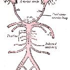
The Circle of Willis is an arterial polygon (heptagon) formed as the internal carotid and vertebral systems anastomose around the optic chiasm and infundibulum of the pituitary stalk in the suprasellar cistern. This communicating pathway allows equalization of blood-flow between the two sides of the brain, and permits anastomotic circulation, should a part of the circulation be occluded.
Gross anatomy
Vessels comprising the circle of Willis include:
- anterior circulation
- left and right internal carotid arteries (ICA)
- horizontal (A1) segments of the left and right anterior cerebral arteries (ACA)
- single anterior communicating artery (ACOM)
- posterior circulation
- left and right posterior communicating arteries (PCOM) (although some consider the PCOM to be anterior circulation)
- horizontal (P1) segments of left and right posterior cerebral arteries (PCA)
- single basilar artery (tip)
The basilar artery divides at the upper border of the pons to form the left and right PCAs. From each ICA, a PCOM arises at the anterior perforated substance and runs back through the interpeduncular cistern to join the ipsilateral PCA. Each ICA also gives off an ACA. The ACAs are united by the ACOM, a small vessel that runs in the chiasmatic cistern (below the rostrum of the corpus callosum), to complete the circle.
Branches of the circle of Willis also supply the optic chiasm and tracts, infundibulum, hypothalamus and other structures at base of brain:
- medial lenticulostriate arteries (from the A1 segment of ACA)
- thalamoperforating and thalamogeniculate arteries (from the basilar tip, proximal PCAs, and PCOMs)
- perforating branches (from the ACOM)
Variant anatomy
A complete circle of Willis (in which no component is absent or hypoplastic) is only seen in 20-25% of individuals. Posterior circulation anomalies are more common than anterior circulation variants and are seen in nearly 50% of anatomical specimens.
Common variants
- hypoplasia of one or both PCOM ~30% (range 25-34%)
- hypoplastic/absent A1 segment of ACA ~15% (range 10-15%)
- absent or fenestrated ACOM ~12.5% (range 10-15%)
- origin of PCA from the ICA with absent/hypoplastic P1 segment (fetal PCA) ~20% (range 17-25%)
- infundibular dilatation of the PCOM origin ~10% (range 5-15%)
Congenital absence of one or both ICAs may occur but is rare. If one ICA is absent, intrasellar intercarotid communicating arteries are common and there is a high incidence of associated aneurysms.
Carotid-vertebrobasilar anastomoses
- persistent primitive trigeminal artery (PPTA), seen in 0.1-0.6% of cerebral angiograms; bilateral PTAs extremely rare. PTA arises where the ICA exits the carotid canal to enter the cavernous sinus; it then runs posterolaterally along the trigeminal nerve (41%), or crosses over or through the dorsum sellae (59%) before joining the basilar artery. Usually associated with a small PCOM and vertebrals, as well as a hypoplastic basilar caudal to the anastomosis of PTA (increased incidence of AVMs and aneurysms).
- persistent primitive hypoglossal artery (PPHA), seen in 0.027-0.26% of cerebral angiograms. It courses through the hypoglossal canal, parallel to the nerve, connecting the cervical ICA with the basilar artery. When present, it is functionally a single artery that supplies the brainstem and cerebellum (often associated with aneurysms).
- persistent otic (acoustic) artery
- persistent proatlantal artery
See persistent carotid-vertebrobasilar anastomoses.
History and etymology
It is named after the English physician Thomas Willis (1621–1675), who first described the anatomy of his circle in 1664 in his book "Cerebri anatome: cui accessit nervorum descriptio et usus" (The Anatomy of the Brain and Nerves). He called his discovery the "circulus arteriosus cerebri".
He was also responsible for the numbering of the cranial nerves, still used to this day.
He died in 1675 from pleurisy and his remains are buried at Westminster Abbey in London, UK.
Siehe auch:
- Arteria carotis interna
- Arteria vertebralis
- Cerebellum
- Arteria cerebri anterior
- Varianten Circulus Willisi
- Arteria cerebri posterior
- Arteria basilaris
- Hypothalamus
- Arteria trigemina
- Anatomie Hirnstamm
- medial lenticulostriate arteries
- Hypophysenstiel
- Arteria communicans anterior
- foetal PCOM
- posterior communicating arteries (PCOM)
und weiter:

 Assoziationen und Differentialdiagnosen zu Circulus Willisi:


