Moyamoya-Muster

Preschooler
with cafe au lait spots. AP view of a 3D maximum intensity projection of an MRA of the neck with contrast (above) and AP (below left) and inferior (below right) views of a 3D maximum intensity projection of an MRA of the brain without contrast shows diffuse hypoplasia of the left internal carotid artery. There is stenosis and occlusion of the cavernous and supraclinoid segments of the left internal carotid artery and then there is a thin left M1 segment without evidence of collaterals in the basal ganglia. Additionally, aneurysms are noted in the left A1 and A2 segments.The diagnosis was Moya Moya syndrome in a patient with neurofibromatosis type I.

Neuroimaging
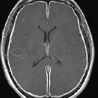
assessment in Down syndrome: a pictorial review. Axial FLAIR image in a 12-year-old girl with Down syndrome and moyamoya pattern depicting the classical ivy sign, corresponding to areas of absence of normal FLAIR suppression of CSF within cortical sulci. This sign probably represents slow flow in leptomeningeal collaterals. The exam was performed without general anesthesia

Neuroimaging
assessment in Down syndrome: a pictorial review. Cerebral angiography in a 46-year-old Down syndrome patient with moyamoya pattern. a Frontal view, right internal carotid artery injection, showing occlusion of the right anterior cerebral artery, with a network of collateral vessels (moyamoya vessels) in the supra-sellar cistern with reconstitution of the anterior cerebral artery arterial flow distally. b Lateral view, left internal carotid artery injection, showing severe narrowing of this artery, which ends on the cavernous segment, where the arterial flow diverges to a meningohypophyseal trunk with anastomosis with the ophthalmic artery and dural branches of the middle meningeal artery, forming a network of collateral capillaries, typical of moyamoya syndrome

Sickle cell
disease cerebral vasculopathy. Sickle cell vasculopathy with occlusion of internal carotid arteries and moyamoya pattern supported by posterior circulation from vertebrobasilar system. Notice patent bilateral superficial temporal artery synangiosis.

Moyamoya
syndrome • Moyamoya disease - Ganzer Fall bei Radiopaedia

Moyamoya
syndrome • Moyamoya phenomenon - Ganzer Fall bei Radiopaedia

Moyamoya
syndrome • Moya moya phenomenon - Ganzer Fall bei Radiopaedia

Sickle cell
disease (cerebral manifestations) • Moyamoya syndrome with background sickle cell disease - Ganzer Fall bei Radiopaedia

Moyamoya
syndrome • Moyamoya syndrome associated with Down syndrome - Ganzer Fall bei Radiopaedia

Moyamoya
syndrome • Moyamoya syndrome associated with type I diabetes mellitus - Ganzer Fall bei Radiopaedia
Moyamoya syndrome, also termed the moyamoya pattern or phenomenon, is due to numerous conditions that can cause arterial occlusion of the circle of Willis, with resultant collaterals, and appearances reminiscent of moyamoya disease. These conditions include :
- vessel wall abnormalities
- phakomatoses
- connective tissue disorders
- blood dyscrasias
- sickle cell disease
- essential thrombocytopenia
- polycythemia rubra vera
- aplastic anemia
- Fanconi anemia
- infection
- less common causes
- Graves disease
- Down syndrome
- ulcerative colitis (UC)
- Apert syndrome
- oral contraceptive use
Siehe auch:
- Tuberöse Sklerose
- Neurofibromatose Typ 1
- Down-Syndrom
- Fibromuskuläre Dysplasie
- Morbus Basedow
- Circulus Willisi
- Sichelzellenanämie
- Colitis ulcerosa
- Apert-Syndrom
- Moyamoya-Erkrankung
- Atherosklerose
- bakterielle Meningitis
- tuberkulöse Meningitis
- Leptospirose
und weiter:

 Assoziationen und Differentialdiagnosen zu Moyamoya-Muster:


