Radiation-induced cerebral vasculopathy


Radiation-induced cerebral vasculopathy or cranial arteritis encompasses a complex and broad range of effects on the intra- and extracranial vessels resulting from injury from radiation exposure. Manifestations can include hemorrhages and ischemic strokes, cavernoma and capillary telangiectasias, and large vessel stenosis mimicking moyamoya pattern.
Clinical presentation
Clinical symptoms are broad and depend on the underlying vasculopathy. Radiation-induced telangiectasia and microbleeds may be asymptomatic. However, patients may present with focal neurological deficits resulting from both hemorrhagic and ischemic stroke, lacunar lesions, vascular occlusive disease including moyamoya pattern, vascular malformations, and radiation-induced necrosis.
The likelihood of complications depends on individual patient factors, including age, and the specifics of the type and dose of radiation.
Pathology
A number of processes occur in vessels following radiation-induced injury and are dependent on dose, however, the dominant earliest phenomenon is endothelial damage. This not only results in disruption of the blood-brain-barrier but also may result in local thrombosis and hemorrhage .
Following repair, vessels tend to be dilated and demonstrate endothelial proliferation, basement membrane thickening and adventitial fibrosis .
Relatively low doses tend to result in the development of capillary telangiectasias, whereas high doses have more profound effects including blood vessel wall necrosis .
Biopsy and pathological analysis of radiation-induced telangiectasias have demonstrated deposition of perivascular hemosiderin and hemorrhage adjacent to capillary-sized telangiectasia (pre-existing dilated channels rather than proliferating vascular neoplasm) .
Veins are affected less than arteries and capillaries .
Radiographic features
The imaging features are different depending on the manifestation and are thus discussed separately, however, it should be noted that unlike sporadic cases the distribution of changes is usually confined to the radiation field.
- cavernoma
- capillary telangiectasia
- large vessel stenosis (moyamoya pattern)
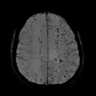
- intracranial hemorrhage (including cerebral microhemorrhages)
- ischemic stroke
- radionecrosis (not purely a vasculopathy)
MRI
Vessel wall imaging can demonstrate vessel wall thickening and enhancement of the large cerebral arteries .
Treatment and prognosis
Treatment is directed at the underlying specific disease and ranges from expectant (for telangiectasias) to intensive standard management of cerebral hemorrhages and ischemic strokes.
Elective angioplasty and stenting of significant large arterial stenoses may also be appropriate for high-grade lesions .

