intracerebral hemorrhage

Intracranial
hemorrhage • Evolution of MRI signal characteristics of intracranial hemorrhage (diagram) - Ganzer Fall bei Radiopaedia

Intracerebral
hemorrhage • Hyperacute intracerebral hemorrhage on MRI and CT - Ganzer Fall bei Radiopaedia

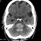
CT-scan of a
frontal intracerebral hemorrhage on the right (left image side) as a contre coup. There is dens suture material visible left occipital in the skin.

Hypertensive
intracerebral hemorrhage • Hypertensive basal ganglial bleed - Ganzer Fall bei Radiopaedia

Intracerebral
hemorrhage • Hemorrhagic infarcts - vertebral artery occlusion - Ganzer Fall bei Radiopaedia

Hypertensive
intracerebral hemorrhage • Cerebellar hemorrhage - Ganzer Fall bei Radiopaedia

Hypertensive
intracerebral hemorrhage • Hypertensive intracerebral hemorrhage - Ganzer Fall bei Radiopaedia

Hypertensive
intracerebral hemorrhage • Hypertensive hemorrhage - Ganzer Fall bei Radiopaedia

Intracerebral
hemorrhage • Hemorrhagic cerebral infarction - Ganzer Fall bei Radiopaedia

Intracerebral
hemorrhage • Intracerebral hemorrhage - Ganzer Fall bei Radiopaedia

Intracerebral
hemorrhage • Cerebral hematoma - Ganzer Fall bei Radiopaedia

Intracerebral
hemorrhage • Subacute intracranial hemorrhage - Ganzer Fall bei Radiopaedia

Intracerebral
hemorrhage • Coagulopathy related intracerebral hemorrhage - Ganzer Fall bei Radiopaedia

Intracerebral
hemorrhage • Traumatic intracerebral hemorrhage - Ganzer Fall bei Radiopaedia

Intracerebral
hemorrhage • Intracerebral hemorrhage - Ganzer Fall bei Radiopaedia

Magnetic
resonance imaging of arterial stroke mimics: a pictorial review. A 48-year-old woman with intracerebral haemorrhage presenting with left hemiplegia and left homonymous hemianopsia. DWI shows heterogeneous signal abnormalities involving the right lenticular nucleus on b1000 (a), apparent diffusion coefficient (ADC) (b) and fluid-attenuated inversion recovery (FLAIR) (c). The lesion appears as a peripheral hypointensity on T2 with gradient recalled echo weighted imaging (T2-GRE) (d, arrows)

Intracerebral
hemorrhage • Intracerebral hemorrhage - spot sign - Ganzer Fall bei Radiopaedia

Blood -Fluid
Level in a Spontaneous Intraparenchymal hematoma. Cut at basal ganglia shows haematoma with very minimal peri-hematomal edema

Intracerebral
hemorrhage • Intracerebral hemorrhage (warfarinised) - Ganzer Fall bei Radiopaedia

COVID-19
neurological manifestations: correlation of cerebral MRI imaging and lung imaging—Observational study. A 50-year-old male presented with sever continuous headache during COVID infection, expressive aphasia. MRI revealed right occipital well defined intra axial space occupying lesion. a Axial T1 WI showed area of peripheral high SI with central area of iso intense signal intensity. b Axial FLAIR show area of peripheral edema (c) axial T2WI showed area of relative low SI with peripheral edema (d) ADC map and (e) DWI show area of peripheral restriction. Finding correlates with subacute intraparenchymal hematoma. f CT lung showed multifocal GGO and consolidation, very typical COVID abnormalities (CORAD-V)

Blood -Fluid
Level in a Spontaneous Intraparenchymal hematoma. Cut at body of ventricle shows haematoma with blood-fluid level

Newborn with
congenital diaphragmatic hernia on ECMOCoronal and sagittal US of the brain shows a large, round echogenic lesion in the left parietal lobe.The diagnosis was intracerebral hemorrhage on ECMO.

Newborn who
is on ECMO after cardiac surgeryCoronal US of the brain (below) shows echogenic material in right subdural space. Coronal and sagittal US of the brain (above) shows a right parietal round mixed echogenicity lesion.The diagnosis was subdural hematoma on ECMO and intracerebral hemorrhage on ECMO.

Comparison of
conventional gradient echo image (GRE) with TE=20 ms (left) and susceptibility weighted image (SWI) with TE=40 ms (right) collected at 1.5 Tesla. Subject shows hemorrhage from trauma.

Comparison of
conventional gradient echo T2*-weighted image (left, TE=20ms), susceptibility weighted image (SWI) and SWI phase image (center and right, respectively, TE=40ms) at 1.5 Tesla. Low signal foci in cerebral amyloid angiopathy (CAA) is shown.

MRI-scan (T1)
of a frontal intracerebral hemorrhage on the right (left image side) as a contre coup.

MRI-scan (T2*
Haem) of a frontal intracerebral hemorrhage on the right (left image side) as a contre coup.

Einige Tage
alter kortikaler Hirninfarkt rechts hochfrontal mit geringer Hämorrhagie. MRT T1 koronar. Man erkennt das Infarktareal leicht hypointens und darin schlierenartig eine Hyperintensität, die dem ausgetretenen Blut entspricht (Pfeil).
An intracerebral hemorrhage, or intraparenchymal cerebral hemorrhage, is a subset of an intracranial hemorrhage and encompasses a number of entities that have in common the acute accumulation of blood within the parenchyma of the brain. The etiology, epidemiology, treatment and prognosis vary widely depending on the type of hemorrhage, and as such, these are discussed separately.
They are most often broadly divided according to whether they are spontaneous (primary) or due to an underlying lesion (secondary), and then further divided according to etiology and/or location.
- vascular malformation
- cerebral venous thrombosis
- tumor (primary or secondary)
Practical points
With any intracerebral hemorrhage the following points should be included in a report as they have prognostic implications :
- location
- size/volume
- the ABC/2 formula is widely used, but there may be more accurate formulas (e.g. 2.5ABC/6, SH/2) and analyzes available, some of which, however, may require the addition of specific software to the standard PACS tools
- shape (irregular vs regular)
- density (homogeneous vs heterogeneous)
- presence/absence of substantial surrounding edema that may indicate an underlying tumor
- presence/absence of intraventricular hemorrhage
- presence/absence of hydrocephalus
- when CT angiography is performed, the presence/absence of the CTA spot sign or a vascular malformation
Video
Siehe auch:
- Altersbestimmung Blutung MRT
- Intrakranielle Blutung
- Ponsblutung
- zerebrale Mikroblutungen
- Einblutung in die Basalganglien
- Kleinhirnblutung
- Lobärblutung
- Hirnblutung mit Ventrikeleinbruch
- subpiale Blutung
- intracranial hemorrhage evaluation with mri
- zerebraler Mittellinienshift
- Einteilung Hirnblutung
- hypertensive haemorrhage
- haemorrhagic venous infarct
und weiter:
- zerebrale Verkalkungen
- Zerebrale Amyloidangiopathie
- Einklemmung
- Ischämischer Schlaganfall
- intraventrikuläre Blutung
- neuroradiologisches Curriculum
- kapilläre Teleangiektasien des ZNS
- intrakranielle Thrombektomie
- hypertensive Hirnblutung
- Coup-Contre-coup-Mechanismus
- Schlaganfall
- systemic hypertension
- intermediary injury
- hyperdense intracerebral lesions
- zerebrale Kontusionsblutung
- paediatric intraventricular haemorrhage
- intracranial haemorrhage with fluid-fluid level
- Hirnstammblutung
- Carotis-Sinus-cavernosus-Fistel
- Hirnblutung und Infarkt beim Neugeborenen
- cerebral abscesses secondary to contusions
- intracerebral haemorrhage (warfarinised)
- intrazerebrale Blutung bei Amyloidangiopathie
- zerebrale Blutung beim Neugeborenen
- akute intrazerebrale Blutung
- cerebral lobar haematoma secondary to amyloid angiopathy
- Hämosiderinablagerungen nach intrazerebraler Blutung
- Thalamusblutung
- Hirnherniation
- Subarachnoidalblutung nach Schlangenbiss


 Assoziationen und Differentialdiagnosen zu Intrazerebrale Blutung: