phakomatoses

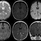
Sturge-Weber-Syndrom
bei einem Säugling: Asymmetrie mit kleinerer linker Hemisphäre. Vertiefte Sulci links mit Verschmälerung der Gyri. Nach Kontrastmittelgabe (unten Mitte und rechtes Bild) pathologische Anreicherung der pialen Gefäße über der linken Hemisphäre. Keine Diffusionsstörung.

This image
is part of a series which can be scrolled interactively with the mousewheel or mouse dragging. This is done by using Template:Imagestack. The series is found in the category Neurofibromatosis type I - orbital dysplasia - MRI - case 001. Orbitadysplasie bei Neurofibromatose Typ 1. MRT - T2w axial


Tuberöse
Sklerose in der MRT: Subependymales Knötchen im Seitenventrikel in der Nähe des Foramen Monroi mit deutlicher Kontrastmittelaufnahme. Die Entwicklung eines Riesenzelltumors kann nur im Verlauf (Größenzunahme oder Änderung der Kontrastmittelaufnahme) vermutet werden.

Phakomatoses
• Sturge-Weber syndrome - Ganzer Fall bei Radiopaedia
Phakomatoses are a group of neurocutaneous disorders characterized by the involvement of structures that arise from the embryonic ectoderm (thus central nervous system, skin, and eyes). Other organs may also be involved.
Pathology
As a group, they are characterized by widespread abnormalities often with characteristic appearances. There are around 30 phakomatoses of which the most important ones are listed here:
- neurofibromatosis type I (NF I)
- neurofibromatosis type II (NF II)
- tuberous sclerosis
- Sturge-Weber syndrome
- von Hippel-Lindau syndrome
Siehe auch:
- Tuberöse Sklerose
- Neurofibromatose Typ 1
- Sturge-Weber-Krabbe-Syndrom
- Gorlin-Goltz-Syndrom
- Morbus Hippel-Lindau
- Neurofibromatose Typ 2
- Cowden-Syndrom
- PHACES syndrome
- progressive faziale Hemiatrophie
- Haberland-Syndrom
- Louis-Bar-Syndrom
und weiter:

 Assoziationen und Differentialdiagnosen zu Phakomatosen:


