Gorlin-Goltz-Syndrom

Masses of
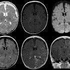
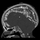
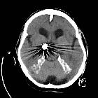
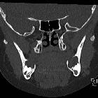
developmental and genetic origin affecting the paediatric craniofacial skeleton. Typical manifestations of nevoid basal cell carcinoma syndrome (NBCCS) in a 16-year-old boy. a Orthopantomography (OPT) shows cystic lesions of the mandible and maxilla (arrows), with unilocular and multilocular pattern and smooth or scalloped borders associated with displaced and unerupted permanent teeth. b Coronal CT scan (bone window) shows ectopic calcifications of the falx cerebri and tentorium cerebelli (arrows) and spotted meningeal calcifications (arrowheads). Brain MRI reveals a cavum veli interpositi on axial T2 (asterisk in c) and coronal contrast-enhanced T1 (asterisk in d) and also vermian dysgenesis (arrowheads in d)
Gorlin-Goltz
syndrome • Gorlin-Goltz syndrome - Ganzer Fall bei Radiopaedia

Gorlin-Goltz
syndrome • Gorlin syndrome - Ganzer Fall bei Radiopaedia
Gorlin-Goltz
syndrome • Gorlin-Goltz syndrome - Ganzer Fall bei Radiopaedia
Gorlin-Goltz
syndrome • Gorlin-Goltz syndrome - incidental posterior communicating aneurysm - Ganzer Fall bei Radiopaedia
Gorlin-Goltz
syndrome • Odontogenic keratocysts - Ganzer Fall bei Radiopaedia
Gorlin-Goltz
syndrome • Gorlin-Goltz syndrome - Ganzer Fall bei Radiopaedia

School ager
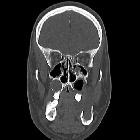
with a bump on the right side of the face. Axial (above), coronal (below left) and sagittal (below right) CT without contrast of the face shows a lytic, expansile lesion in the right maxilla displacing the second right premolar tooth anterolateraly. There was no bone destruction or associated soft tissue mass.The diagnosis was an odontogenic keratocyst in a patient with Gorlin syndrome.

Radiolucent
lesions of the mandible: a pattern-based approach to diagnosis. Basal cell nevus syndrome. a OPG. Multiple mandibular and maxillary KCOTs (asterisks) associated with impacted teeth. b Intraoperative view showing cheese-like material within the angulo-mandibular lesion (arrow). c Surgical specimen showing the KCOT (asterisk) and the associated tooth (thin long arrow). d Histology (haematoxylin-eosin stain, original magnification 40×): corrugated (dashed arrows) parakeratinised epithelium with distinct basal columnar cells with inverted polarity (arrows) and flat connective tissue interface
 nicht verwechseln mit: Goltz-Gorlin-Syndrom
Gorlin-Goltz-Syndrom
Basalzellkarzinome in Kombination mit multiplen Kieferzysten und RippenanomalienSiehe auch:
- Pectus carinatum
- Pectus excavatum
- Medulloblastom
- Cherubismus
- Hypertelorismus
- Gabelrippe
- frontal bossing
- Syndaktylie
- Makrozephalie
- Lippen-Kiefer-Gaumen-Spalte
- Halbwirbel
- Keratozystischer odontogener Tumor
- Kolobom
- Fibrom des Ovars
- Mikrophthalmus
- Dysgenesie des Corpus callosum
- Phakomatosen
- kardiales Fibrom
- lytische Läsionen der Mandibula
- Läsionen der Mandibula
- Trichoepitheliom
- shortened 4th metacarpal bones
und weiter:
- angeborene Wirbelanomalien
- Café-au-lait-Fleck
- subkutane Verkalkungen
- orbital hypertelorism
- Basaliom
- conditions involving skin and bone
- Verkürzung Metakarpale vier oder fünf
- Greig-Syndrom
- short metacarpals (mnemonic)
- Goltz-Gorlin-Syndrom
- keratocystic odentogenic tumour
- Zyste Maxilla
- keratocyst
- osteogenic jaw cyst
- diffuse Duraverkalkungen


 Assoziationen und Differentialdiagnosen zu Gorlin-Goltz-Syndrom: