posterior cerebral artery

Middle
cerebral artery (MCA) infarct • Vascular territories of the lateral cerebral cortex (illustration) - Ganzer Fall bei Radiopaedia

The brain and
arteries at base of the brain. Circle of Willis is formed near center. The temporal pole of the cerebrum and a portion of the cerebellar hemisphere have been removed on the right side. Inferior aspect (viewed from below).

Posterior
cerebral artery • Accessory PCA arising from the terminal ICA - Ganzer Fall bei Radiopaedia

Middle
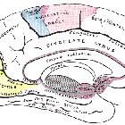
cerebral artery (MCA) infarct • Cerebral vascular territories in the midline (illustration) - Ganzer Fall bei Radiopaedia
Middle
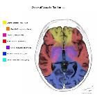
cerebral artery (MCA) infarct • Cerebral vascular territories (illustration) - Ganzer Fall bei Radiopaedia

Anterior
cerebral artery • Common variants of the circle of Willis (illustrations) - Ganzer Fall bei Radiopaedia

Anterior
inferior cerebellar artery • Brainstem arterial territories (diagrams) - Ganzer Fall bei Radiopaedia

Posterior
cerebral artery • Posterior cerebral artery segments - Ganzer Fall bei Radiopaedia

Posterior
cerebral artery • PCA infarction - Ganzer Fall bei Radiopaedia
The posterior cerebral arteries (PCA) are the terminal branches of the basilar artery and supply the occipital lobes and posteromedial temporal lobes.
Summary
- origin: terminal branches of the basilar artery
- course: from basilar towards occiput
- main branches
- supply: occipital lobes and posteromedial temporal lobes
Gross anatomy
The PCA is divided into four segments:
- P1: pre-communicating segment
- originates at the termination of the basilar artery
- terminates to the posterior communicating artery (PCOM), within the interpeduncular cistern
- P2: post-communicating segment
- from the PCOM around the midbrain
- P2A (anterior): sub-segment courses through the crural cistern
- P2P (posterior or ambient): sub-segment courses through the ambient cistern
- terminates as it enters the quadrigeminal cistern
- from the PCOM around the midbrain
- P3: quadrigeminal segment
- courses posteromedially through the quadrigeminal cistern
- terminates as it enters sulci of the occipital lobe
- P4: cortical segment
- within the sulci of the occipital lobe
- e.g. calcarine artery, within the calcarine fissure
Branches
- posterior communicating artery
- collicular (quadrigeminal) artery
- choroidal branches (from P2)
- perforators
- anterior thalamoperforator (from PCOM)
- posterior thalamoperforator (from P1)
- thalamogeniculate perforator (from P2)
- peduncular perforator (from P2)
- circumflex (long and short)
- cortical branches
- temporal branches
- anterior temporal artery
- posterior temporal artery
- lateral occipital artery
- anterior inferior temporal artery
- middle inferior temporal artery
- posterior inferior temporal artery
- medial occipital artery
- calcarine artery
- parieto-occipital artery
- splenial artery
- temporal branches
Supply
The posterior cerebral artery curls around the cerebral peduncle and passes above the tentorium to supply the posteromedial surface of the temporal lobe and the occipital lobe. The visual cortex responsible for the contralateral field of vision lies in its territory. The macular part of the visual cortex often receives a dual blood supply from the PCA and the MCA, which explains the "macular sparing" phenomenon in some patients following a PCA infarct.
Variant anatomy
- fetal origin of PCA: unilateral incidence 10%, bilateral incidence 8%
- PCA fenestration: rare
- duplicated PCA: rare, fetal origin and normal origin on same side
Siehe auch:
- Circulus Willisi
- Arteria basilaris
- Cisterna quadrigeminalis
- Percheron-Arterie
- posterior communicating artery (PCOM)
- Sulcus calcarinus
- Normvarianten Arteria cerebri posterior
- calcarine artery
- foetal PCOM
- temporal lobes
und weiter:
- Thalamus
- calcarine fissure
- Precuneus
- fetal PCOM
- basal vein of Rosenthal
- embryonaler Versorgungstyp Arteria cerebri posterior
- neuroradiologisches Curriculum
- arterielle Versorgungsgebiete des Gehirns
- posterior cerebral circulation
- Arteria cerebelli superior
- posterior choroidal artery
- Infarkt der Arteria cerebri posterior
- Nervus oculomotorius
- yasargil classification
- Gefäßversorgung Gehirn
- medial posterior choroidal artery
- lateral posterior choroidal artery
- Segmente Arteria cerebri posterior
- Grenzzoneninfarkt
- posterior choroidal artery stroke
- Migräne
- foetal posterior comminucating artery


 Assoziationen und Differentialdiagnosen zu Arteria cerebri posterior: