bakterielle Zystitis

Catheter-related
lower urinary tract infection complicated by mural bladder abscess. Portal phase enhanced images showed persistent uniform urothelial hyperenhancement (thin arrow) consistent with acute infectious cystitis; peripheral rim enhancement of the abscess collection (arrowheads) abutting the bladder dome.

Catheter-related
lower urinary tract infection complicated by mural bladder abscess. Excretory phase acquisition showed opacified bladder lumen, uniformly thickened bladder wall (*), enhanced periphery of the abscess (arrowheads) abutting the bladder dome. No appreciable communication between lumen and abscess.

Catheter-related
lower urinary tract infection complicated by mural bladder abscess. Preliminary unenhanced acquisition showed contracted urinary bladder with indwelling Foley catheter (short arrows), circumferential mural thickening (*), several centimetric calcific calculi.

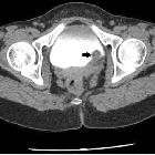
Multidetector
computed tomography evaluation of bladder lesions. A 62-year-old female patient with acute bacterial cystitis. Axial pre-contrast (a) and contrast-enhanced (b) CT images showing diffuse enhancement of the mucosal surface with wall thickening of the bladder


 Assoziationen und Differentialdiagnosen zu bakterielle Zystitis: