cystitis glandularis


Cystitis glandularis is a proliferative disorder of the urinary bladder in which there is glandular metaplasia of the transitional cells lining the urinary bladder. This entity is closely related to cystitis cystica, with which it commonly co-exists. It is a relatively common chronic reactive inflammatory disorders that occur in the setting of chronic irritation of the bladder mucosa, often identified on biopsies and cystectomies. It usually occurs in the trigone of the urinary bladder.
Epidemiology
Cystitis cystica is seen in a variety of patients, who all have chronic bladder inflammation as a uniting feature. The underlying causes include:
- chronic bladder outlet obstruction
- chronic infection
- bladder calculi
Pathology
Chronic irritation from infection, calculi or even tumors results in metaplasia of the urothelium, which proliferates into buds, which grow down into the connective tissue beneath the epithelium in the lamina propria. In the case of cystitis glandularis, the buds then differentiate into mucin-producing goblet cells (whereas in cystitis cystica they differentiate into fluid-filled cysts). In most cases, examples of both conditions can be identified histologically.
There are two main types of cystitis glandularis, non-mucinous and mucinous (intestinal). There could also be focal or diffuse involvement of the bladder. The intestinal subtype of cystitis glandularis and diffuse lesions have been proposed to be a precursor of adenocarcinoma. However, certain authors have considered cystitis glandularis to be a chronic and quiescent histologic lesion without any clinical significance.
Radiographic features
Fluoroscopy
On IVP, there is a lobulated outline of the urinary bladder with a nodular filling defect within.
Ultrasound
Often seen as focal polypoidal wall thickening of the urinary bladder in the region of the trigone.
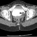
CT
Hypervascular polypoid masses within the urinary bladder, best delineated on delayed (urography) phase.
MRI
- T1: may be seen as low signal polypoidal lesion
- T2:
- low signal lesion with central branching hyperintensity
- central hyperintensity enhances on contrast administration and represents vascular stalk
Treatment and prognosis
Treatment consists of removing the source of irritation and surgical excision of the area of inflammation or cystectomy in rare severe cases. An association with adenocarcinoma of the bladder has been described, and thus these patients should be monitored.
Differential diagnosis
- transitional cell carcinoma (urinary bladder) - An intact muscle layer is seen in cystitis cystica and cystitis glandularis. This usually requires an MRI in order to better distinguish the bladder walls layers.
Siehe auch:
und weiter:


 Assoziationen und Differentialdiagnosen zu cystitis glandularis:

