Zystitis

Emphysematöse
Cystitis in der Computertomografie ohne und mit retrograder Kontrastmittelfüllung der Blase über einen Blasenkatheter

Catheter-related
lower urinary tract infection complicated by mural bladder abscess. Portal phase enhanced images showed persistent uniform urothelial hyperenhancement (thin arrow) consistent with acute infectious cystitis; peripheral rim enhancement of the abscess collection (arrowheads) abutting the bladder dome.

Catheter-related
lower urinary tract infection complicated by mural bladder abscess. Excretory phase acquisition showed opacified bladder lumen, uniformly thickened bladder wall (*), enhanced periphery of the abscess (arrowheads) abutting the bladder dome. No appreciable communication between lumen and abscess.

Catheter-related
lower urinary tract infection complicated by mural bladder abscess. Preliminary unenhanced acquisition showed contracted urinary bladder with indwelling Foley catheter (short arrows), circumferential mural thickening (*), several centimetric calcific calculi.

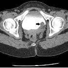
Multidetector
computed tomography evaluation of bladder lesions. A 62-year-old female patient with acute bacterial cystitis. Axial pre-contrast (a) and contrast-enhanced (b) CT images showing diffuse enhancement of the mucosal surface with wall thickening of the bladder
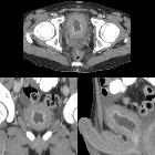
Teenager with
new hematuria who is being treated with cyclophosphamide for lymphoma. Axial (above), coronal (below left) and sagittal (below right) CT with contrast of the abdomen shows diffuse thickening of the bladder wall and hyperemia of the bladder mucosa. The diagnosis was cyclophosphamide induced cystitis.
School ager
with gross hematuria who is post bone marrow transplant and has received cyclophosphamide. Transverse (above) and sagittal (below) US of the pelvis shows an extremely thick-walled bladder containing echogenic debris and clots.The diagnosis was hemorrhagic cystitis due to cyclophosphamide therapy.


 Assoziationen und Differentialdiagnosen zu Zystitis: