Mediastinalshift

Mediastinalshift
bei asymmetrischer emphysematischer Überblähung. Rechts Röntgen, links CT zu einem anderen Zeitpunkt damals mit Infiltrat rechts.

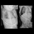
Rotationsfehler
in der Bettlunge nach links mit Unterlappenatelektase links: Der (echte) Mediastinalshift nach links wird durch den Rotationsfehler übertrieben.

Reexpansionsödem
der Lunge links nach Entlastung eines Pleuraergusses. Im linken Bild großer Pleuraerguss links mit Mediastinalshift. Im rechten Bild Z.n. Anlage einer Pleuradrainage, welche sich auf den lateralen Recessus projiziert. Im Mittelfeld flaue Ödemzone nach Reexpansion. Mediastinaler Shift wieder ausgeglichen.

Fremdkörperaspiration:
Die linke Lunge ist durch die Ventilwirkung des Fremdkörpers im linken Hauptbronchus deutlich überbläht. Entsprechend Mediastinalshift nach rechts. Der Fremdkörper konnte bronchoskopisch entfernt werden.
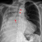
Mediastinalshift
nach rechts bei subtotaler Atelektase der rechten Lunge bei Bronchialkarzinom (Pfeil). Die Pfeilspitzen zeigen auf die nach rechts verschobene Luftröhre.

Tension
pneumothorax • Tension pneumothorax - Ganzer Fall bei Radiopaedia


Fremdkörperaspiration
(Erdnuss oder Apfelstück?) mit Ventilmechanismus und Überblähung der rechten Lunge. Deutlicher Mediastinalshift nach links.

Pleurakuppenschwiele
und Pleurosis calcarea nach Tuberkulose. Durch die narbige Schrumpfung ist das Volumen der Thoraxhälfte rechts deutlich vermindert, sodass es zu einem deutlichen Mediastinalshift kommt.

Spontanpneumothorax
rechts (links im Bild) mit leichtem Mediastinalshift im Sinne eines Spannungspneumothorax. Beachte auch den basalen Spiegel. 2 Jahre zuvor Spontanpneumothorax auf der anderen Seite.

Mäßiger
(bzgl. der Spannungskomponente) spontaner Spannungspneumothorax links mit etwas Mediastinalshift nach rechts. Man erkennt gut die kollabierten Lungenlappen.


Ausgedehnte
kongenitale Zwerchfellhernie links: Linker Hemithorax verschattet. Magenblase und Spitze der Magensonde oberhalb des erwarteten Zwerchfellniveaus. Herz und Thymus nach rechts verlagert. Rechte Lunge klein und nur geringer Anteil belüftet.

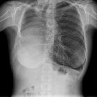
Mediastinalshift
nach rechts bei großem Pleuraerguss links: Beachte den Verlauf der Trachea.


Tension
pneumothorax • Tension pneumothorax (serial imaging) - Ganzer Fall bei Radiopaedia

Tension
pneumothorax • Tension pneumothorax - Ganzer Fall bei Radiopaedia

Tension
pneumothorax • Neonatal pneumothorax - tension - Ganzer Fall bei Radiopaedia

Tension
pneumothorax • Tension pneumothorax - Ganzer Fall bei Radiopaedia

Tension
pneumothorax • Tension pneumothorax mimic due to right lower lobe hypoplasia - Ganzer Fall bei Radiopaedia

Tension
pneumothorax • Tension pneumothorax - Ganzer Fall bei Radiopaedia
Premature
newborn after nasogastric tube placement. Initial CXR AP (left) shows the course of the nasogastric tube to project over the right lung and the tip of the nasogastric tube projects over the liver and is probably in the right costophrenic sulcus. The remaining tubes and lines are in appropriate position. CXR AP obtained after removal of the nasogastric tube (right) shows a large amount of air in the right pleural space and there is mediastinal shift to the left.


 Assoziationen und Differentialdiagnosen zu Mediastinalshift: