laryngotracheales Trauma


laryngotracheales Trauma
tracheobronchiale Verletzungen Radiopaedia • CC-by-nc-sa 3.0 • de
Tracheobronchial injury is a serious but uncommon manifestation of chest trauma. It is usually a fatal injury with only a small percentage of patients making it to hospital. Given the magnitude of force required to injure the major airways, there are often multiple chest injuries and other body regions affected.
Pathology
The cervical trachea is the most injured site in penetrating injury , whereas injury to the distal trachea (within 2.5cm of the carina) is seen most commonly following blunt chest trauma . The site of injury can be identified in 70% of patients with CT .
Radiographic features
Plain radiograph
- subcutaneous emphysema in the chest and/or neck
- pneumothorax
- pneumomediastinum
- endotracheal tube balloon
- overinflation
- abnormal shape
- herniation into the airway defect
CT
Features include :
- disruption of the tracheal or bronchial cartilage rings
- irregularity of the airway wall
- focal thickening of the tracheal or bronchial wall
- laryngeal disruption
- massive pneumomediastinum (sometimes despite appropriate pleural space decompression with intercostal catheters)
- fallen lung sign (first described in 1970 ): rare
Complications
If left untreated, late complications can occur such as :
- recurrent infection (pneumonia)
- bronchial or tracheal stenosis
- bronchiectasis
Differential diagnosis
- tracheal diverticulum: usually on the right and at the level of the superior thoracic aperture
Larynxverletzungen Radiopaedia • CC-by-nc-sa 3.0 • de
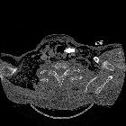
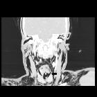
Laryngeal trauma is uncommon in the setting of external blunt or penetrating trauma. The larynx may also be injured internally, for example during endotracheal intubation.
Clinical presentation
Symptoms include hoarseness, laryngeal pain, dyspnea, and/or dysphagia. Also, stridor, hemoptysis, subcutaneous emphysema and tenderness/deformity of the larynx skeleton may be present. Laryngeal trauma in a significant number of patients may be clinically occult .
Pathology
Etiology
The larynx may be injured in a number of ways :
- external blunt: motor vehicle accidents, "clothesline" accidents, strangulation, falls, sporting injuries
- external penetrating: stabbing, shooting
- internal blunt: iatrogenic during endotracheal intubation or fibroendoscopic examination
Location
The following laryngeal structures may be injured in isolation or combination :
- laryngeal edema, hematoma and/or mucosal lacerations
- thyroid and/or cricoid cartilage fracture
- laryngeal rupture
- cricotracheal separation
- cervical tracheal injury
The hyoid bone, although technically not part of the larynx, can also be fractured.
Associations
Other injuries are common :
- base of skull or facial fractures (~20%)
- cervical spine fractures (~10%)
- thoracic and abdominal injuries
Radiographic features
CT is the modality of choice. Review of airway obstruction includes evaluation of edema, hematoma, foreign body or displaced fractures.
CT review for larynx function should also include close inspection for lacerations especially to the vocal cord and anterior commissure, and arytenoid cartilage dislocation or avulsion.
Treatment and prognosis
The key goals of management are:
- maintaining airway: preserve life
- restoring function to larynx: voice quality
Blunt trauma has a worse prognosis than penetrating trauma with increased length of stay and short-term mortality (40% vs 20%) respectively .
Complications
Complications include :
- airway stenosis
- respiratory tract infection
- aspiration
- tracheo-esophageal fistula
Practical points
- detection of cartilage fractures
- evaluation of the subglottic and anterior commissure regions which are poorly visualized on endoscopy
Siehe auch:

 Assoziationen und Differentialdiagnosen zu laryngotracheales Trauma:

