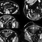
Riesenpseudodivertikel des Kolons


Giant colonic diverticula, also referred to as giant colonic pseudodiverticula, are an uncommon form of presentation of colonic diverticulosis and are characterized by large diverticular masses, usually filled with stool and gas, that communicate with the colonic lumen.
Terminology
Although the great majority of cases are not true colonic diverticula (cf. pseudodiverticula or chronic contained perforation of the colon), this article will follow the majority of publications and the way the condition is commonly referred to in daily practice, which means grouping all types under the umbrella term of giant colonic diverticula.
Clinical presentation
The most common presentation is abdominal pain. Other presenting symptoms include constipation, abdominal masses, vomiting, diarrhea, and rectal bleeding .
Pathology
The majority (>90%) arise from the sigmoid colon , but they can occur in any part of the colon. They are divided into three different types :
- the second most common type (~20% of cases)
- the mucosa herniates through the muscular wall and becomes progressively enlarged by colonic gas via a ball-valve type mechanism
- diverticular wall is made of remnants of both the muscularis mucosa and muscularis propria, which are permeated by chronic granulation tissue
- majority of cases (~70% of cases)
- result from perforation and formation of a persistent abscess cavity that remains in communication with the colonic lumen
- it can also be understood as a chronic contained colonic perforation
- diverticular wall is made of dense fibrous tissue containing both chronic inflammatory cells and foreign body giant cells
- rare (~10% of cases)
- contain all the colonic wall layers: serosa, muscularis, submucosa, and mucosa
- believed to represent a duplication cyst that has communication with the colonic lumen
Radiographic features
Imaging plays an important role in the diagnosis of giant colonic diverticula as they might not be identified on colonoscopy studies due to a small ostium .
Plain radiograph
Abdominal x-rays may show large gas-filled cysts with a gas-fluid level .
CT
CT is the best imaging modality for diagnosis given that colonoscopies are rarely carried out due to the risk of perforation . CT shows the diverticulum as a gas, fluid or feces-filled cavity, sometimes with calcification in the case of chronic inflammation. There is usually no contrast enhancement unless there is active inflammation .
Treatment and prognosis
The definitive treatment for a giant diverticulum is surgery .
Complications
- common
- perforation
- abscess formation
- less common
- intestinal volvulus
- intestinal obstruction
- lower gastrointestinal bleeding
- lymphoma
- adenocarcinoma arising in the diverticulum
Siehe auch:
- Pneumoperitoneum
- Sigmadivertikulose
- Duplikationszysten gastrointestinal
- Dünndarmdivertikel
- Kolondivertikulitis
- Kolondivertikulose
- Fußballzeichen (Pneumoperitoneum)
und weiter:

 Assoziationen und Differentialdiagnosen zu riesiges Sigmadivertikel:


