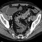
colonic diverticulosis


Colonic diverticulosis (plural diverticuloses) refers to the presence of multiple diverticula. It is quite distinct from diverticulitis which describes inflammation and infection of one or multiple diverticula.
Epidemiology
Diverticulosis is very common in westernised countries and is typically found in older individuals. At 40 years of age, ~5% of the population have diverticula; at 60 years ~30%, increasing to 50-80% by the age of 80 .
Clinical presentation
The vast majority of people with diverticulosis are asymptomatic. Nevertheless, since prevalence is high, diverticula are a common reason for presentation and admission to hospital.
Patients complain of intermittent left sided abdominal pain and frequent constipation. Symptomatic presenting features of diverticular disease (i.e. presentation of complicated diverticulosis) include:
- diverticulitis
- lower gastrointestinal hemorrhage ("diverticular hemorrhage")
Pathology
Colonic diverticula are almost all false diverticula: mucosa herniating through a defect in the muscularis and covered by overlying serosa (where present). This herniation typically occurs where nutrient arteries enter the colon , and therefore, are more common on the mesenteric side of the colon.
They are thought to relate to increased intraluminal pressure which may be a result of low volume stool. Increased pressure is thought to be related to chronic constipation and straining. The colon is shortened and hypertrophied (myochosis coli).
There is also an increased incidence of diverticula amongst patients with connective tissue disorders, e.g. Ehlers-Danlos syndrome, Marfan syndrome, scleroderma .
Colonic diverticula are most common in the sigmoid colon and, to a lesser extent, in the descending colon. The entire colon can be affected, however, with 15% of patients having right-sided diverticula . In patients from Asia, right-sided diverticula are more common, and can either be single or multiple .
The entity of giant colonic diverticulum is uncommon and usually arises from the sigmoid colon .
Radiographic features
Diverticula range in size from a few millimeters to a few centimeters .
Barium enema
Both single and double contrast barium enemas are able to demonstrate diverticula as barium-filled outpouchings.
When seen 'en face' they can look similar to polyps but can be distinguished by the presence of contrast pooling within the diverticulum and forming a meniscus. Even if seen on a recumbent overhead film, the two can usually be separated. (see differential below)
Ultrasound
Diverticula can be visualized as gas-filled outpouchings from the colon, especially if use of linear array probe is allowed by patient anatomy.
CT
Diverticula are usually outlined by gas. The colon may be thickened and shortened. On CT VR colonoscopy, they are seen as complete rings delineating the neck.
Treatment and prognosis
The majority of patients remain asymptomatic throughout life and no treatment is required. A high fiber diet may reduce the incidence of diverticula and rate of complications . Treatment is usually reserved for diverticulitis or diverticular hemorrhage.
Differential diagnosis
The differential for uncomplicated diverticula is small, but they do need to be differentiated from polyps on barium studies and CT virtual colonoscopy because of potentially similar appearances. Additionally, when significant mural thickening is present due to muscular hypertrophy, diverticulosis need to be distinguished from colorectal carcinoma given the treatment implications.
Polyps
- on double contrast barium enema, diverticula must be distinguished from polyps, but this is usually trivial
- when seen 'en face' they can look similar to polyps but can be distinguished by the presence of pooling contrast within the diverticulum and forming a meniscus
- even if seen on a recumbent overhead film, the two can usually be separated; contrast will often outline the pedicle of the polyp and fade out peripherally, whereas the rounded meniscus pooling in the diverticulum with fade centrally
- on CT VR colonoscopy, diverticula will be seen as complete rings delineating the neck, whereas polyps will have an incomplete ring
Colorectal carcinoma
- rolled/heaped-up shoulders
- short segment
- nodal enlargement
- for more information, see colorectal carcinoma
Siehe auch:
- Duodenaldivertikel
- Sigmadivertikulitis
- Kolorektales Karzinom
- Sigmadivertikulose
- Colon sigmoideum
- Kolonkontrasteinlauf
- Divertikulose
- Kolondivertikulitis
- AWMF - Leitlinie Divertikelkrankheit - Divertikulitis
und weiter:
- Pneumatosis intestinalis
- myochosis coli
- Dünndarmdivertikel
- Kolondivertikulose
- Divertikelblutung
- colovaginal fistula
- intramurale Gaseinschlüsse
- Stenose Divertikulitis
- colonic adenomatous polyps and diverticular disease
- diverticulum of the caecum
- Divertikel des Gastrointestinaltraktes
- Saint-Trias
- Divertikel
- Rektumdivertikel
- Riesenpseudodivertikel des Kolons

 Assoziationen und Differentialdiagnosen zu Kolondivertikulose:


