Strahlenpneumonitis


Radiation pneumonitis is the acute manifestation of radiation-induced lung disease and is relatively common following radiotherapy for chest wall or intrathoracic malignancies.
This article does not deal with the changes seen in the late phase. Please refer to the article on radiation-induced lung disease for a general discussion and radiation-induced pulmonary fibrosis for specific discussion of these late changes.
Epidemiology
For a discussion of the epidemiology of radiation-induced lung disease please refer to the parent article: radiation-induced lung disease.
Clinical presentation
Radiation pneumonitis typically occurs between 4 and 12 weeks following completion of radiotherapy course, although they may be seen as early as one week, especially in patients receiving a high total dose and/or also having received chemotherapy .
Symptoms typically include :
- cough
- dyspnea (exertional or at rest)
- low-grade fever
- chest discomfort
- pleuritic pain
Pathology
The lungs are the most sensitive organ when irradiating the chest, and are the major dose-limiting factor. Radiation pneumonitis reflects the acute response of the lung to radiation and includes :
- loss of type I pneumocytes
- increased capillary permeability resulting in
- interstitial and alveolar edema
- ingress of inflammatory cells into the alveolar spaces
When changes are seen in the non-irradiated lung, immune-mediated lymphocytic alveolitis has been postulated as the underlying cause .
Radiographic features
Although changes in the lung are usually confined to the irradiated port, changes in the remainder of the lung may also on occasion be seen .
Plain radiograph
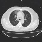
Chest x-ray changes are non-specific but confined to the irradiation port, with airspace opacities being most common. Pleural effusions or atelectasis are also sometimes seen .
CT
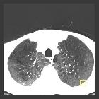
CT is not only better able to delineate parenchymal changes, but often demonstrates changes localized to the irradiated field, making the diagnosis easier. With stereotactic ablative radiotherapy the shape of the irradiated field will not have straight edges or conform to the traditional conventional radiotherapy portals. As such it may be less obviously artificial in shape .
In cases of early or subtle radiation-induced pneumonitis, areas of ground-glass opacity may be evident on CT despite a normal chest x-ray .
The two most common findings are ground-glass opacities and/or airspace consolidation . Unusual patterns of airspace opacities include :
- halo sign: lung consolidation surrounded by ground-glass opacities
- reversed halo sign: ground-glass opacities surrounded by a crescentic or circumferential area of consolidation
Additional features that are sometimes seen include :
- nodule-like pattern
- first few months
- tend to resolve or coalesce
- tree-in-bud appearances
- the differential with superimposed infection should be considered
- septal thickening may occur later with the alveolar opacities producing a “crazy paving” pattern
- ipsilateral pleural effusion
- seen within 6 months after the completion date
- usually small
- can persist for months
- atelectasis
- pulmonary necrosis: rare
- chronic pleural effusion
- usually small
- smooth local pleural thickening can be seen
FDG-PET
FDG-PET performed soon after completion of radiotherapy often demonstrates increased metabolic activity in both lungs, especially in a peripheral distribution. Only a minority of patient go on to develop clinically or CT evident pneumonitis .
FDG avidity in the treated area is usually present in late phases of radiation pneumonitis (3 to 9 months after treatment completion) due to the presence of residual inflammation and, therefore, PET-CT is of equivocal clinical value in this period .
Treatment and prognosis
Steroids can reduce the severity of acute radiation pneumonitis. Depending on the degree of injury changes may be mild and spontaneously resolve or progress adult respiratory distress syndrome with a high rate of mortality . Chronically radiation fibrosis may occur .
Differential diagnosis
If a clear demarcation conforming to the irradiation port is seen then there is little difficulty in making the diagnosis, especially when a history of chest radiotherapy is known. In cases where the distribution is atypical the differential depends on the dominant feature:
A knowledge of the time course of the changes concerning radiotherapy, total dose administered, administration of chemotherapy, and shape of the portal used can all have a significant impact on the differential, and thus should be sought if the referring clinician has not provided them .
Siehe auch:
- Milchglasverschattungen
- strahleninduzierte Lungenerkrankung
- differential of chronic airspace opacities
und weiter:

 Assoziationen und Differentialdiagnosen zu Strahlenpneumonitis:

