Tree-in-bud sign (lung)


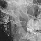
Tree-in-bud sign or pattern describes the CT appearance of multiple areas of centrilobular nodules with a linear branching pattern. Although initially described in patients with endobronchial tuberculosis, it is now recognized in a large number of conditions.
Pathology
Pathogenesis
Simply put, the tree-in-bud pattern can be seen with two main sites of disease :
- distal airways
- distal pulmonary vasculature
More specifically, the pattern can be manifest because of the following disease processes, often in combination:
- airway-centered:
- bronchioles filled with pus or inflammatory exudate
- bronchiolitis: thickening of bronchiolar walls and bronchovascular bundle
- bronchiectasis/bronchiolectasis with mucus plugging
- e.g. cystic fibrosis
- bronchovascular interstitial infiltration
- e.g. sarcoidosis, lymphoma, leukemia
- vascular-centered
- tumor emboli to centrilobular arteries (or carcinomatous endarteritis)
- e.g. breast cancer, stomach cancer
- granulomatous response to excipient material in intravenous drug abusers
- e.g. intravenous talcosis or microcrystalline cellulose in crushed oral tablets (excipient lung disease)
- tumor emboli to centrilobular arteries (or carcinomatous endarteritis)
Etiology
While the tree-in-bud appearance usually represents an endobronchial spread of infection, given the proximity of small pulmonary arteries and small airways (sharing branching morphology in the bronchovascular bundle), a rarer cause of the tree-in-bud sign is infiltration of the small pulmonary arteries/arterioles or axial interstitium .
Causes include:
- infective bronchiolitis
- congenital
- connective tissue disorders
- bronchial
- neoplastic (i.e. carcinomatous endarteritis or bronchovascular interstitial infiltration )
- bronchioloalveolar cell carcinoma
- distant metastatic disease (e.g. breast, liver, ovary, prostate, kidney)
- primary pulmonary lymphoma
- chronic lymphocytic leukemia
- periarterial granulomatous
Radiographic features
Tree-in-bud sign is not generally visible on plain radiographs . It is usually visible on standard CT, however, it is best seen on HRCT chest. Typically the centrilobular nodules are 2-4 mm in diameter and peripheral, within 5 mm of the pleural surface. The connection to opacified or thickened branching structures extends proximally (representing the dilated and opacified bronchioles or inflamed arterioles) .
Practical points
- using maximum intensity projection (MIP) can facilitate detection of particularly the centrilobular nodules
- identification of the tree-in-bud sign should urge you to
- look for further imaging findings e.g. thickening of the bronchial wall, narrowing of bronchi, bronchiectasis, consolidation, cavitation, necrotic lymphadenopathy
- determine the location (with gravitational or lower lobe predominance favoring aspiration)
- scrutinise patient history, including appropriate exposure history, as this may aid in determining the most likely diagnosis
Siehe auch:
- Bronchiektasen
- Rheumatoide Arthritis
- Bronchiolitis obliterans
- pulmonale Tuberkulose
- Allergische bronchopulmonale Aspergillose
- Kartagener-Syndrom
- In Situ Adenokarzinom der Lunge
- zystische Fibrose
- Bronchiolitis
- Sjögren-Syndrom
- Aspirationspneumonie
- Pneumocystis jiroveci Pneumonie
- primäre ciliäre Dyskinesie
- zentrilobuläre Lungennoduli
- aspergillus
- diffuse panbronchiolitis
- chronic lymphocytic leukaemia
- follikuläre Bronchiolitis
- Nichttuberkulöse Mycobakteriose
- Neoplasien der Mamma
- primäres pulmonales Lymphom
- Mycobacterium avium
- endobronchial tuberculosis
- (MAIC)
und weiter:

 Assoziationen und Differentialdiagnosen zu tree in bud-Muster:


