fetal schizencephaly

Fetal
schizencephaly • Fetal schizencephaly - Ganzer Fall bei Radiopaedia
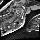
Fetal MRI of
post-clastic schizencephaly. Fetal brain shows bilateral open lip schizencephaly with asymmetry of the lateral ventricles at the schizencephaly defects. T2 hypo rim is visible as sign of post-haemorrhagic tissue disruption.

Fetal MRI of
post-clastic schizencephaly. Fetal brain shows bilateral open lip schizencephaly with asymmetry of the lateral ventricles at the schizencephaly defects. T2 hypo rim is visible as sign of post-haemorrhagic tissue disruption.

Fetal MRI of
post-clastic schizencephaly. Fetal brain shows bilateral open lip schizencephaly with asymmetry of the lateral ventricles at the schizencephaly defects. T2 hypo rim is visible as sign of post-haemorrhagic tissue disruption.
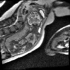
Fetal MRI of
post-clastic schizencephaly. Fetal brain shows bilateral open lip schizencephaly with asymmetry of the lateral ventricles at the schizencephaly defects. T2 hypo rim is visible as sign of post-haemorrhagic tissue disruption.
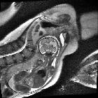
Fetal MRI of
post-clastic schizencephaly. Fetal brain shows bilateral open lip schizencephaly with asymmetry of the lateral ventricles at the schizencephaly defects. T2 hypo rim is visible as sign of post-haemorrhagic tissue disruption.
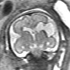
Fetal MRI of
post-clastic schizencephaly. Fetal brain shows bilateral open lip schizencephaly with asymmetry of the lateral ventricles at the schizencephaly defects. T2 hypo rim is visible as sign of post-haemorrhagic tissue disruption.
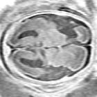
Fetal MRI of
post-clastic schizencephaly. Fetal brain shows bilateral open lip schizencephaly with asymmetry of the lateral ventricles at the schizencephaly defects.
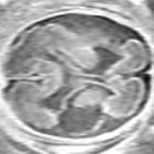
Fetal MRI of
post-clastic schizencephaly. Fetal brain shows bilateral open lip schizencephaly with asymmetry of the lateral ventricles at the schizencephaly defects.

Fetal MRI of
post-clastic schizencephaly. Fetal brain shows bilateral open lip schizencephaly with asymmetry of the lateral ventricles at the schizencephaly defects.
Fetal schizencephaly refers to schizencephaly diagnosed in utero. Usually only open lips types can be diagnosed antenatally.
Radiographic features
Antenatal ultrasound
- may show a unilateral or bilateral defect extending from the pial surface to the ventricular wall
- there may be other features such as
- absent cavum septum pellucidum
- occasional fetal hydrocephalus
Fetal MRI
- fetal MRI is performed to confirm the cleft is grey matter lined which distinguishes this entity from porencephalic cyst
- it is more sensitive at detecting close lip schizencephaly than ultrasound
- it is also useful to confirm the presence of associated anomalies
Practical points
Similar abnormalities in the posterior fossa are called cerebellar clefts, and may occur simultaneously with fetal schizencephaly.
Siehe auch:

 Assoziationen und Differentialdiagnosen zu fetale Schizenzephalie:

