venöse Aneurysmen

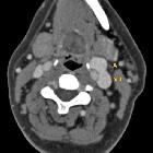
Internal
jugular vein aneurysm. Sagittal sectional CT image demonstrating aneurysm of left IJV
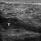
Post-thrombotic
aneurysmal dilatation of the hypogastric vein. 5 years earlier, longitudinal (a) and transverse (b,c) ultrasound images showed chronic deep venous thrombosis (T) of the right lower limb, from the popliteal to the common femoral vein.
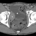
Post-thrombotic
aneurysmal dilatation of the hypogastric vein. The venous phase acquisition (d-h) showed the lesion (*) to enhance homogeneously and synchronously with venous vessels, consistent with aneurysmal dilatation of the proximal hypogastric vein. Urinary bladder (+).
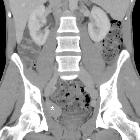
Post-thrombotic
aneurysmal dilatation of the hypogastric vein. Coronal (f) and sagittal (g,h) reconstructions confirmed aneurysmal dilatation (*) of the right hypogastric vein without signs of thrombosis. No abnormal dilatation, compression or thrombosis of the inferior cava, common and external iliac veins.
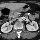
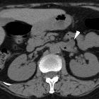
Asymptomatic
left renal vein aneurysm with thrombosis. Multiplanar reformatted images (b..d) from portal venous phase acquisition depicted the lesion (arrowheads) as a saccular outpouching of the left renal vein (*), with internal nonenhancing tissue (thin arrows) representing intraluminal thrombus.


 Assoziationen und Differentialdiagnosen zu venöse Aneurysmen: