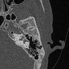
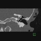
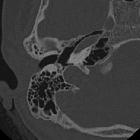
fenestrale Otosklerose


Otosclerosis
• Fenestral otosclerosis - Ganzer Fall bei Radiopaedia

Role of MRI
as first-line modality in the detection of previously undiagnosed otosclerosis: a single tertiary institute experience. A 64-year-old male patient with heavy bilateral SNHL (case 7). Axial CISS image (a) shows curvilinear T2 hyperintensities (arrow) surrounding the left cochlea. Subsequent axial HRCT image (b) confirms extensive very hypodense plaques (arrows) in the left pericochlear region, likely long-standing disease. Axial CISS image (c) at a lower level also shows a small right IAC diverticulum (dashed arrow), confirmed on subsequent axial HRCT image (d) as an ‘ear-like’ outpouching anterior to the right IAC (dashed arrow). Also note the large plaque in the right FA region (short arrow)

Role of MRI
as first-line modality in the detection of previously undiagnosed otosclerosis: a single tertiary institute experience. A 41-year-old male patient with left-sided SNHL (case 4). Axial T1W MR image (a) shows intermediate T1 signal in the right FA (arrow). Contrast-enhanced T1W MR image (b) shows moderate enhancement in bilateral FA regions (arrows) and mild enhancement in the right pericochlear region (dashed arrow). Pericochlear enhancement was not seen on the left. Subsequent axial HRCT images (c, d) confirm bilateral fenestral otosclerotic plaques (short white arrows) and pericochlear plaques (black long arrows). The right RW is occluded (curved arrow)

Role of MRI
as first-line modality in the detection of previously undiagnosed otosclerosis: a single tertiary institute experience. A 57-year-old female patient with left-sided SNHL (case 3). Axial T1W MR image (a) shows intermediate T1 signal in in bilateral pericochlear regions (arrows). Contrast-enhanced axial T1W MR image (b) and contrast-enhanced coronal T1W MR image (c) show ring-like enhancement in bilateral pericochlear regions (arrows). Contrast-enhanced coronal T1W MR image (d) also shows patchy enhancement surrounding the SCC bilaterally (dashed arrows). Subsequent coronal HRCT images (e, f) confirm otosclerotic plaques adjacent to bilateral lateral SCC (short white arrow), superior SCC (dashed arrow) and in bilateral pericochlear regions (long black arrows)

Role of MRI
as first-line modality in the detection of previously undiagnosed otosclerosis: a single tertiary institute experience. A 46-year-old male patient with right-sided MHL (case 8). Axial T1W MR image (a) shows intermediate T1 signal around the basal turn of the right cochlea (thin arrow) and in the left FA region (thick arrow). Axial T1W MR image at a lower level (b) shows intermediate T1 signal around the basal turn of the left cochlea (thin arrow) and another separate focus of intermediate signal in the left pericochlear region (thick arrow). Contrast-enhanced axial T1W image (c) shows moderate post-contrast enhancement in bilateral FA regions (arrows) and mild enhancement around the basal turns of cochleae (dashed arrows). Contrast-enhanced axial T1W image at a lower level (d) shows mild enhancement around the basal turn of left cochlea (dashed arrow) and another focus of enhancement in the left pericochlear region (arrow). Subsequent axial HRCT images (e, f) confirm bilateral fenestral otosclerotic plaques (short white arrows) and pericochlear plaques (black long arrows)

Role of MRI
as first-line modality in the detection of previously undiagnosed otosclerosis: a single tertiary institute experience. A 28-year-old male patient with left-sided SNHL (case 2). This was the case where otosclerosis was missed on first read MRI. Retrospective evaluation of axial T1W MR images (a, b) shows very subtle intermediate T1 signal in bilateral FA regions (arrows). Contrast-enhanced axial T1W images (c, d) show subtle focal post-contrast enhancement in bilateral FA regions (arrows). Axial HRCT images (e, f) show bilateral fenestral otosclerotic plaques (arrows). There was no pericochlear disease seen on HRCT

Role of MRI
as first-line modality in the detection of previously undiagnosed otosclerosis: a single tertiary institute experience. A 43-year-old female patient with right-sided SNHL (case 1). Axial T1W MR images (a, b) show subtle intermediate T1 signal in bilateral FA regions (arrows). Contrast-enhanced axial T1W images (c, d) show moderate post-contrast enhancement in bilateral FA regions (arrows) and mild post-contrast enhancement in bilateral pericochlear regions (dashed arrows). Subsequent axial HRCT images (e, f) confirm bilateral fenestral otosclerotic plaques (short white arrow) and pericochlear plaques (black long arrows)

Otosclerosis
• Otospongiosis (fenestral and retrofenestral) - Ganzer Fall bei Radiopaedia

Otosclerosis
• Fenestral otosclerosis - Ganzer Fall bei Radiopaedia

Otosclerosis
• Otospongiosis - Ganzer Fall bei Radiopaedia

Otosclerosis
• Otosclerosis - Ganzer Fall bei Radiopaedia


Conductive
hearing loss • Fenestral otosclerosis - Ganzer Fall bei Radiopaedia

Otosclerosis
• Fenestral otosclerosis - Ganzer Fall bei Radiopaedia

Otosclerosis
• Otosclerosis - fenestral - Ganzer Fall bei Radiopaedia

Otosclerosis
• Bilateral otosclerosis with left stapes prosthesis - Ganzer Fall bei Radiopaedia


CT
contribution in otosclerosis. Hypodensity pericochlear and in front of acoustic meatus


CT
contribution in otosclerosis. Hypodense plaque around the cochlea

CT
contribution in otosclerosis. Hypodense plaque at the edges of the oval window perish

Pre- and
post-operative imaging of cochlear implants: a pictorial review. A 70-year-old male patient, with progressive SNHL from advanced otosclerosis. HRCT axial (left image) and paracoronal (right image) image show irregular ossifications affecting the cochlea (black arrows). Vestibulum and semicircular canals (white arrows) are not affected
 Assoziationen und Differentialdiagnosen zu fenestrale Otosklerose:


