Tumoren des hinteren Mediastinums


Unicentric
Castleman disease. Posteroanterior chest X-ray demonstrates a left paravertebral mass, of smooth contours, that follows the left paraspinal line.

Primary
posterior mediastinal yolk sac tumour in a two-year-old girl. Heterogeneous mass with the extension into the pleural cavity and inferiorly into the liver. Aerated lung and small amount of collapsed lung are at the superior aspect of the mass.

Primary
posterior mediastinal yolk sac tumour in a two-year-old girl. heterogeneous echogenic mass (arrow) in the right pleural cavity surrounded by pleural fluid (star).

Primary
posterior mediastinal yolk sac tumour in a two-year-old girl. Heterogeneous attenuation mass causing the left shift of the heart (arrow).

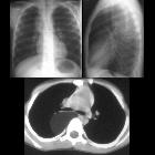
School ager
with cough and fever. CXR PA and lateral (above) shows a right sided chest mass in the posterior mediastinum. Axial CT with contrast of the chest (below) shows a homogeneous mass, of fat density, with a few septations, in the right posterior mediastinum causing some anterior displacement of the right mainstem bronchus.The diagnosis was lipoblastoma in the posterior mediastinum.
Tumoren des hinteren Mediastinums
Siehe auch:
- Hiatushernie
- Chordom
- Lymphom
- Neuroblastom
- extramedulläre Hämatopoese
- Schwannom
- Bochdalek'sche Hernie
- Phäochromozytom
- maligner peripherer Nervenscheidentumor (MPNST)
- Neurofibrom
- neuroenterische Zyste
- Duplikationszyste des Ösophagus
- benignes Ganglioneurom
- Lymphadenopathie
- neurogenic tumours
- neuroblastic tumours
und weiter:
- Osteomyelofibrose
- Morbus Castleman
- mediastinale Raumforderungen
- hinteres Mediastinum
- posterior mediastinal mass in the exam
- Liposarkom des Mediastinums
- pleomorphic liposarcoma of the posterior mediastinum
- primary liposarcoma of the posterior mediastinum
- Yolk sac tumour arising within the posterior mediastinum

 Assoziationen und Differentialdiagnosen zu Tumoren des hinteren Mediastinums:


