nasogastric tube

Premature
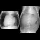
newborn with abdominal distension after nasogastric tube placementSupine AXR (above) shows increased lucency in the upper abdomen. The cross-table lateral AXR (below) shows air between the anterior abdominal wall and the bowel. The nasogastric tube follows a very straight course and does not curve towards the stomach. Note on both views that the tip of the nasogastric tube does not project over the stomach. US performed subsequently showed the tip of the nasogastric tube was in the retroperitoneum and not in the stomach.The diagnosis was perforation of the esophagus during nasogastric tube placement. In the operating room the pneumoperitoneum was found to be due to an ileal perforation.

Newborn with
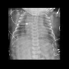
distended abdomen after nasogastric tube placementSupine AXR (left) shows a large amount of air within the abdomen and air outlining both sides of bowel wall (Rigler’s sign) in the right lower quadrant. Supine AXR taken later after pulling back of the nasogastric tube out of the stomach shows visualization of the falciform ligament over the spine (American football sign)The diagnosis was pneumoperitoneum due to gastric perforation during nasogastric tube placement.

Premature
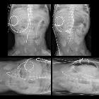
newborn after nasogastric tube placementSupine and left lateral decubitus AXR (left) show a nasogastric tube with its tip deep in the pelvis without evidence of free air. Supine AXR taken after pulling the nasogastric tube back into the stomach (above right) shows increased lucency throughout the central abdomen and left lateral decubitus AXR taken at same time (below right) shows air between the abdominal wall and the liver.The diagnosis was pneumoperitoneum due to gastric perforation during nasogastric tube placement which became visible only after the nasogastric tube was pulled out of the hole it had made in the stomach wall.

Newborn with
hypoxic ischemic encephalopathyCXR AP shows a nasogastric tube coursing within the esophagus into the stomach while the twisted wire probe next to it with its tip in the mid-esophagus represents an esophageal temperature probe in appropriate position. The patient is lying upon a cooling blanket while being cooled to treat their hypoxic ischemic encephalopathy.The diagnosis was a patient with hypoxic ischemic encephalopathy with a mimic of a nasogastric tube.
 Assoziationen und Differentialdiagnosen zu Magensonde:

