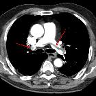
Lungenembolie-CT

Pulmonary
embolism in computertomography. The patient suffered from portal vein thrombosis a year later.

Pulmonary
embolism • Pulmonary embolism - Ganzer Fall bei Radiopaedia

CT pulmonary
angiogram (protocol) • Pulmonary embolism on suboptimal CTPA (spectral low monoE) - Ganzer Fall bei Radiopaedia

CT pulmonary
angiogram (protocol) • Pulmonary embolism (spectral CTPA) - Ganzer Fall bei Radiopaedia

CT pulmonary
angiogram (protocol) • Normal spectral CTPA - Ganzer Fall bei Radiopaedia

CT pulmonary
angiogram (protocol) • Saddle pulmonary embolus - Ganzer Fall bei Radiopaedia

CT pulmonary
angiogram (protocol) • Normal CTPA - Ganzer Fall bei Radiopaedia

Pulmonary
embolism • Pulmonary embolism - Ganzer Fall bei Radiopaedia

Pulmonary
embolism • Pulmonary embolism - Ganzer Fall bei Radiopaedia

Pulmonary
embolism • Pulmonary embolism - Ganzer Fall bei Radiopaedia

Pulmonary
embolism • Pulmonary embolus - Ganzer Fall bei Radiopaedia

Pulmonary
embolism • Extensive acute pulmonary emboli with right heart strain - Ganzer Fall bei Radiopaedia

Pulmonary
embolism • Incidental pulmonary embolism - Ganzer Fall bei Radiopaedia


Pulmonary
embolism • Pulmonary embolism - Ganzer Fall bei Radiopaedia

Pulmonary
embolism • Saddle pulmonary embolism - Ganzer Fall bei Radiopaedia

Pulmonary
embolism • Acute pulmonary embolism - Ganzer Fall bei Radiopaedia
A large
pulmonary embolism at the bifurcation of the pulmonary artery (saddle embolism).


Thorax CT of
a 74-year-old man with a long-standing pulmonary embolism (having lasted 3 months) of the artery of the right lower lobe, secondary to a leg fracture, and with long-standing hemoptysis. It shows the embolism, as well as a pulmonary infarction seen as a reverse halo sign. Further information: Reverse halo sign

This image
is part of a series which can be scrolled interactively with the mousewheel or mouse dragging. This is done by using Template:Imagestack. The series is found in the category Pulmonary embolism - CT - case 001. Reitender Thrombus bei Lungenembolie. Genaugenommen sind es hier mehrere zentrale von links nach rechts reitende Thromben. Die linken Hauptstämme sind weitestgehend okkludiert.

CT pulmonary
angiogram (protocol) • Aortic dissection (CTPA) - Ganzer Fall bei Radiopaedia
Lungenembolie-CT
Computertomographie des Thorax Radiopaedia • CC-by-nc-sa 3.0 • de
Computed tomography (CT) of the chest is a cross-sectional evaluation of the heart, airways, lungs, mediastinum, and associated bones and soft tissues.
Two key methods of image acquisition include:
- standard CT with 5 mm slice thickness for mediastinum and gross evaluation of lungs
- high-resolution CT (HRCT) with thin sections (slice thickness of 0.625 to 1.25 mm) for evaluation of the secondary lobule of the lungs
General indications
Emergencies
- chest trauma: evaluation of contusions, rib fractures, and pneumothorax
- aortic pathologies: dissection, transection
- pulmonary embolism
- post-thoracic surgery complications: mediastinal hematomas, complex pleural collections
Non-emergencies
- evaluation of nodules, hilar, or mediastinal masses identified on a chest radiograph
- diagnosis and staging of lung cancer
- detection of metastasis from known extrathoracic malignancies
- assessment of congenital anomalies of the thoracic great vessels
- characterization of interstitial lung disease (ILDs)
Siehe auch:
- Lungenarterienembolie
- MRI arteria pulmonalis angiography
- Pulmonalisangiographie
- Lungenembolie Szintigraphie
- Verdacht auf Lungenembolie in der Schwangerschaft
und weiter:


 Assoziationen und Differentialdiagnosen zu Lungenembolie-CT:
