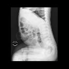
inkarzerierte Nabelhernie

Inkarzerierte
Dünndarmschlinge in einer Nabelhernie bei Patient mit Aszites bei Leberzirrhose: 3 Bilder Sonografie mit der Inkarzeration und computertomografische Voraufnahme mit Darstellung der Hernie und dem massiven Aszites.

Umbilical
hernia • Strangulated umbilical hernia - Ganzer Fall bei Radiopaedia

Umbilical
hernia • Incarcerated umbilical hernia - Ganzer Fall bei Radiopaedia

Umbilical
hernia • Incarcerated umbilical hernia - Ganzer Fall bei Radiopaedia

Small bowel
obstruction • Umbilical hernia causing small bowel obstruction - Ganzer Fall bei Radiopaedia
inkarzerierte Nabelhernie
Umbilikalhernie Radiopaedia • CC-by-nc-sa 3.0 • de
Umbilical hernias (alternative plural: herniae) are the most common ventral abdominal wall hernia and occur in the midline through the umbilicus.
Epidemiology
Ten times more common in females and represent ~5% of all abdominal hernias .
Clinical presentation
Umbilical hernias may present in the midline as a painless or painful mass.
Pathology
Umbilical hernias may be congenital or acquired :
- congenital: physiological herniation through the umbilicus occurs during the 10th week of gestation and congenital umbilical hernias occur when there is incomplete closure of the anterior abdominal wall after the gut returns to the abdominal cavity
- acquired: more common in adults
- risk factors: obesity, multiparity, ascites, large intra-abdominal mass
Umbilical hernias commonly contain fat, mesentery, small and/or large bowel.
Treatment and prognosis
There is a high rate of strangulation and incarceration of bowel and Richter hernias are common. Bowel obstruction is common and can be also be complicated by bowel ischemia.
Differential diagnosis
See also
Siehe auch:

 Assoziationen und Differentialdiagnosen zu inkarzerierte Nabelhernie:

