Tumoren des Peritoneums

Peritoneal
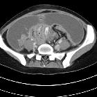
myolipoma. CT images show a large abdominopelvic mass with a smooth contour and a predominantly cystic appearance (10 HU). Areas of fat attenuation (arrows Figs. 1a-1b) and faint enhancing soft-tissue nodules (arrowhead Fig. 1a) are seen.

Peritoneal
myolipoma. CT images show a large abdominopelvic mass with a smooth contour and a predominantly cystic appearance (10 HU). Areas of fat attenuation (arrows Figs. 1a-1b) and faint enhancing soft-tissue nodules (arrowhead Fig. 1a) are seen.

Peritoneal
myolipoma. CT images show a large abdominopelvic mass with a smooth contour and a predominantly cystic appearance (10 HU). Areas of fat attenuation (arrows Figs. 1a-1b) and faint enhancing soft-tissue nodules (arrowhead Fig. 1a) are seen.

Peritoneal
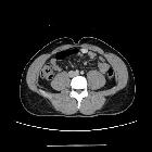
myolipoma. Nodular lesion of soft-tissue attenuation with faint enhancement (arrow).
Tumoren des Peritoneums
Siehe auch:
- Pseudomyxoma peritonei
- Peritonealkarzinose
- peritoneales Mesotheliom
- Tuberkulose des Peritoneums
- primäre peritoneale Neoplasien
- peritoneales Lymphom
- desmoplastischer klein- und rundzelliger Tumor (DSRCT)
- desmoplastischer klein- und rundzelliger Tumor (DSRCT) des Peritoneums
- solitärer fibröser Tumor des Peritoneums
und weiter:


 Assoziationen und Differentialdiagnosen zu Tumoren des Peritoneums:


solitärer
fibröser Tumor des Peritoneums