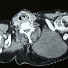
Lipoma vs well-differentiated liposarcoma

Differentiating
atypical lipomatous tumors from lipomas with magnetic resonance imaging: a comparison with MDM2 gene amplification status. Intramuscular lipomatous tumor in a 45-year-old female patient in the left gluteus muscles. (a) The axial T1 weighted image with fat saturation (FS) and (b) the axial T2 weighted image show a well-defined lipomatous tumor which was classified as a lipoma after resection. Another 65-year-old male patient showed an intramuscular lipomatous tumor reaching into the subcutaneous region of the left shoulder. (c) An axial T2 weighted and a (d) coronal T2 weighted image demonstrating pseudo-infiltrative margins of the tumor, which was classified as an ALT after resection

Differentiating
atypical lipomatous tumors from lipomas with magnetic resonance imaging: a comparison with MDM2 gene amplification status. A 41-year-old male patient with a lipomatous tumor (ALT) at the medial left sided thigh showing solid, non-lipomatous components within the tumor on a (a) coronal T1 weighted image with (b) contrast enhancement (> 1/3 of the tumor volume) on a coronal T1 weighted FS image. On the other hand, there is a 45-year-old female patient showing a lipomatous tumor (lipoma) on a (c) sagittal T2 weighted image without (d) contrast enhancement on the coronal T1 weighted FS image in the gluteus region on the left side

Differentiating
atypical lipomatous tumors from lipomas with magnetic resonance imaging: a comparison with MDM2 gene amplification status. Axial T2 weighted images of lipomatous tumors (a) without septa; (b) with thin septa (< 2 mm) and (c) with thick septa (> 2 mm)

Lipoma vs
well-differentiated liposarcoma • Low-grade liposarcoma - Ganzer Fall bei Radiopaedia

Lipoma •
Neck intermuscular lipoma - Ganzer Fall bei Radiopaedia

Lipoma vs
well-differentiated liposarcoma • Soft tissue lipoma - thigh - Ganzer Fall bei Radiopaedia
There a number of features that can help distinguish between lipoma and well-differentiated liposarcoma. This article relates to superficial well-differentiated liposarcomas that typically occur in the extremities, also known as atypical lipomatous tumors, and not retroperitoneal liposarcoma.
Epidemiology
Demographic factors that favor well-differentiated liposarcoma are age >60 years and male sex .
Radiographic features
MRI
MRI can have 100% sensitivity for the diagnosis of well-differentiated liposarcomas, however, has a low positive predictive value due to overlap of imaging features with benign lipoma variants . Features that favor well-differentiated liposarcoma :
- size >10 cm
- percentage fat <75%
- thick or nodular (>2 mm) septa
- nodular/globular areas
- prominent high T2 signal foci
- prominent enhancement
- associated non-adipose mass
- lower limb location
- intramuscular location
Features that favor lipoma :
- size <10 cm
- percentage fat >75%
- no or few thin (<2 mm) septa
- no or minimal enhancement
- no or minimal high T2 signal foci
- subcutaneous location
Siehe auch:

 Assoziationen und Differentialdiagnosen zu Lipom versus Liposarkom:


